Human DLEC/CLEC4C/BDCA-2 Antibody Summary
Phe46-Ile213
Accession # Q8WTT0
Applications
Please Note: Optimal dilutions should be determined by each laboratory for each application. General Protocols are available in the Technical Information section on our website.
Scientific Data
 View Larger
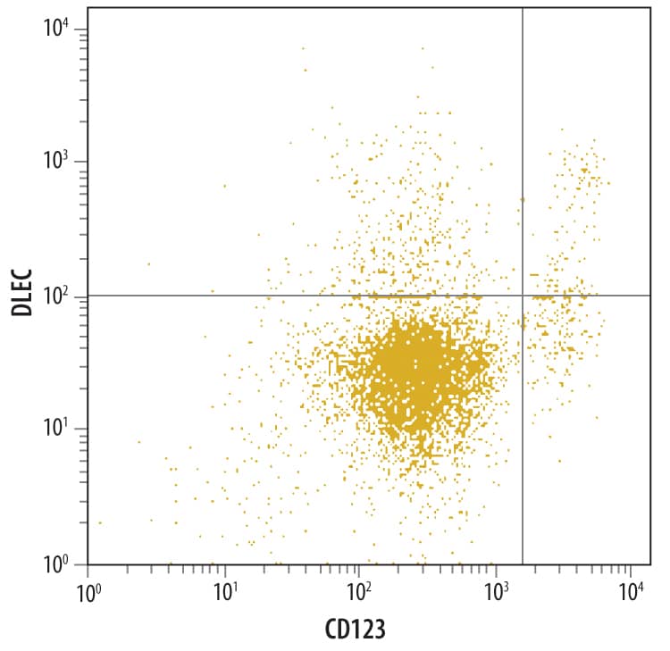
Detection of DLEC/CLEC4C/BDCA‑2 in Human Whole Blood by Flow Cytometry. Human whole blood were stained with Goat Anti- Human DLEC/CLEC4C/BDCA-2 Antigen Affinity-purified Polyclonal Antibody (Catalog # AF1376) followed by Allophycocyanin-conjugated Anti-Goat IgG Secondary Antibody (Catalog # F0108) and Human IL-3 Ra Phycoerythrin-conjugated Monoclonal Antibody (Catalog # FAB301P).Quadrant markers were set based on control antibody staining (Catalog # AB-108-C).
 View Larger
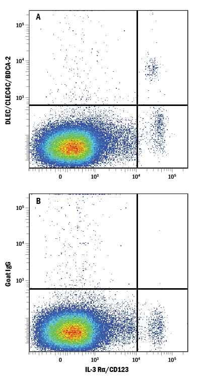
Detection of DLEC/CLEC4C/BDCA‑2 in Human PBMCs by Flow Cytometry. Human peripheral blood mononuclear cells (PBMCs) were stained with Mouse Anti-Human IL-3 Ra/CD123 APC-conjugated Monoclonal Antibody (Catalog # FAB301A) and either (A) Goat Anti-Human DLEC/CLEC4C/BDCA-2 Antigen Affinity-purified Polyclonal Antibody (Catalog # AF1376) or (B) Normal Goat IgG Control (Catalog # AB-108-C) followed by Allophycocyanin-conjugated Anti-Goat IgG Secondary Antibody (Catalog # F0108).
 View Larger
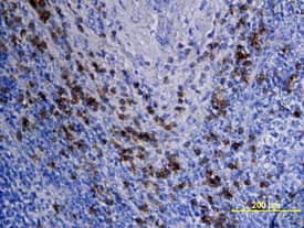
DLEC/CLEC4C/BDCA-2 in Human Lymph Node. DLEC/CLEC4C/BDCA-2 was detected in immersion fixed paraffin-embedded sections of human lymph node using Goat Anti-Human DLEC/CLEC4C/BDCA-2 Antigen Affinity-purified Polyclonal Antibody (Catalog # AF1376) at 15 µg/mL overnight at 4 °C. Tissue was stained using the Anti-Goat HRP-DAB Cell & Tissue Staining Kit (brown; Catalog # CTS008) and counterstained with hematoxylin (blue). View our protocol for Chromogenic IHC Staining of Paraffin-embedded Tissue Sections.
 View Larger
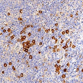
DLEC/CLEC4C/BDCA‑2 in Human Tonsil. DLEC/CLEC4C/BDCA-2 was detected in immersion fixed paraffin-embedded sections of human tonsil using Goat Anti-Human DLEC/CLEC4C/BDCA-2 Antigen Affinity-purified Polyclonal Antibody (Catalog # AF1376) at 3 µg/mL for 1 hour at room temperature followed by incubation with the Anti-Goat IgG VisUCyte™ HRP Polymer Antibody (Catalog # VC004). Tissue was stained using DAB (brown) and counterstained with hematoxylin (blue). Specific staining was localized to lymphocytes. View our protocol for IHC Staining with VisUCyte HRP Polymer Detection Reagents.
Preparation and Storage
- 12 months from date of receipt, -20 to -70 °C as supplied.
- 1 month, 2 to 8 °C under sterile conditions after reconstitution.
- 6 months, -20 to -70 °C under sterile conditions after reconstitution.
Background: DLEC/CLEC4C/BDCA-2
Dendritic cell lectin (DLEC), also known as BDCA-2, CD303, HECL, and CLEC4C/CLECSF11/CLECSF7, is a 38 kDa type II transmembrane protein in the C-type lectin family (1). Mature human DLEC consists of a 21 amino acid (aa) cytoplasmic domain, a 23 aa transmembrane segment, and a 169 aa extracellular domain (ECD) that contains a juxtamembrane neck region and one carbohydrate recognition domain (CRD) (2, 3). Alternate splicing may generate multiple isoforms that lack the transmembrane segment and/or portions of the cytoplasmic, neck, and CRD regions (2-4). An ortholog of human DLEC has not been described in mouse or rat. DLEC expression is restricted to plasmacytoid dendritic cells (pDC) and is downregulated during their maturation (2, 3, 5). pDC play a role in the innate immune response by producing IFN-alpha / beta following exposure to TLR7 and TLR9 agonists such as microbial CpG DNA (3, 5-8). Antibody ligation of DLEC on pDC attenuates the CpG-stimulated production of interferons as well as a Th1 biased response (3, 5-9). DLEC interactions with HIV-1 gp120 and hepatitis B virus soluble antigen may therefore limit the pDC antiviral response (10, 11). Similar to other C-type lectins, DLEC can mediate antigen uptake for MHC loading and presentation to T cells (3, 12). Crosslinking of DLEC on CpG-stimulated pDC inhibits pDC maturation and induces tyrosine phosphorylation on multiple proteins involved in B cell receptor signaling and endocytosis (5, 7, 8). These functions require the association of DLEC with the ITAM-containing Fc epsilon RI gamma chain (7, 8).
- Kanazawa, N. et al. (2004) Immunobiology 209:179.
- Arce, I. et al. (2001) Eur. J. Immunol. 31:2733.
- Dzionek, A. et al. (2001) J. Exp. Med. 194:1823.
- Fernandes, M.J. et al. (2000) Genomics 69:263.
- Wu, P. et al. (2008) Clin. Immunol. 129:40.
- Fanning, S.L. et al. (2006) J. Immunol. 177:5829.
- Rock, J. et al. (2007) Eur. J. Immunol. 37:3564.
- Cao, W. et al. (2007) PloS Biol. 5:e248.
- Riboldi, E. et al. (2009) Immunobiology 214:868.
- Martinelli, E. et al. (2007) Proc. Natl. Acad. Sci. USA 104:3396.
- Xu, Y. et al. (2009) Mol. Immunol. 46:2640.
- Jaehn, P.S. et al. (2008) Eur. J. Immunol. 38:1822.
Product Datasheets
Citations for Human DLEC/CLEC4C/BDCA-2 Antibody
R&D Systems personnel manually curate a database that contains references using R&D Systems products. The data collected includes not only links to publications in PubMed, but also provides information about sample types, species, and experimental conditions.
8
Citations: Showing 1 - 8
Filter your results:
Filter by:
-
The spatial organization of cDC1 with CD8+ T cells is critical for the response to immune checkpoint inhibitors in melanoma patients
Authors: Gobbini, E;Hubert, M;Doffin, AC;Eberhardt, A;Hermet, L;Li, D;Duplouye, P;Ghamry-Barrin, S;Berthet, J;Benboubker, V;Grimont, M;Sakref, C;Perrot, J;Tondeur, G;Harou, O;Lopez, J;Dubois, B;Dalle, S;Caux, C;Caramel, J;Valladeau-Guilemond, J;
Cancer immunology research
Species: Human
Sample Types: Whole Tissue
Applications: Immunohistochemistry -
Characterising plasmacytoid and myeloid AXL+ SIGLEC-6+ dendritic cell functions and their interactions with HIV
Authors: Warner van Dijk, FA;Tong, O;O'Neil, TR;Bertram, KM;Hu, K;Baharlou, H;Vine, EE;Jenns, K;Gosselink, MP;Toh, JW;Papadopoulos, T;Barnouti, L;Jenkins, GJ;Sandercoe, G;Haniffa, M;Sandgren, KJ;Harman, AN;Cunningham, AL;Nasr, N;
PLoS pathogens
Species: Human
Sample Types: Whole Tissue
Applications: Immunohistochemistry -
Guidelines for visualization and analysis of DC in tissues using multiparameter fluorescence microscopy imaging methods
Authors: F Bayerl, DA Bejarano, G Bertacchi, AC Doffin, E Gobbini, M Hubert, L Li, P Meiser, AM Pedde, W Posch, L Rupp, A Schlitzer, M Schmitz, BU Schraml, S Uderhardt, J Valladeau-, D Wilflingse, V Zaderer, JP Böttcher
Oncogene, 2023-01-09;0(0):e2249923.
Species: Human
Sample Types: Whole Tissue
Applications: IHC -
Clinical Significance of Tumor-Infiltrating Conventional and Plasmacytoid Dendritic Cells in Pancreatic Ductal Adenocarcinoma
Authors: I Plesca, I Benešová, C Beer, U Sommer, L Müller, R Wehner, M Heiduk, D Aust, G Baretton, MP Bachmann, A Feldmann, J Weitz, L Seifert, AM Seifert, M Schmitz
Cancers, 2022-02-26;14(5):.
Species: Human
Sample Types: Whole Cells
Applications: IHC -
Immune infiltration phenotypes of prostate adenocarcinoma and their clinical implications
Authors: Zehua Ma, Xiankui Cheng, Ting Yue, Xun Shangguan, Zhixiang Xin, Weiwei Zhang et al.
Cancer Medicine
-
Tissue-specific transcriptional profiling of plasmacytoid dendritic cells reveals a hyperactivated state in chronic SIV infection
Authors: MY Lee, AA Upadhyay, H Walum, CN Chan, RA Dawoud, C Grech, JL Harper, KA Karunakara, SA Nelson, EA Mahar, KL Goss, DG Carnathan, B Cervasi, K Gill, GK Tharp, ER Wonderlich, V Velu, SM Barratt-Bo, M Paiardini, G Silvestri, JD Estes, SE Bosinger
PloS Pathogens, 2021-06-28;17(6):e1009674.
Species: Macaca mulatta (Rhesus Macaque)
Sample Types: Whole Tissue
Applications: IHC -
In situ distribution of HIV-binding CCR5 and C-type lectin receptors in the human endocervical mucosa.
Authors: Hirbod T, Kaldensjo T, Broliden K
PLoS ONE, 2011-09-30;6(9):e25551.
Species: Human
Sample Types: Whole Tissue
Applications: IHC-Fr -
Detection of Intraepithelial and Stromal Langerin and CCR5 Positive Cells in the Human Endometrium: Potential Targets for HIV Infection
Authors: Tove Kaldensjö, Pernilla Petersson, Anna Tolf, Gareth Morgan, Kristina Broliden, Taha Hirbod
PLoS ONE
FAQs
No product specific FAQs exist for this product, however you may
View all Antibody FAQsIsotype Controls
Reconstitution Buffers
Secondary Antibodies
Reviews for Human DLEC/CLEC4C/BDCA-2 Antibody
Average Rating: 5 (Based on 1 Review)
Have you used Human DLEC/CLEC4C/BDCA-2 Antibody?
Submit a review and receive an Amazon gift card.
$25/€18/£15/$25CAN/¥75 Yuan/¥2500 Yen for a review with an image
$10/€7/£6/$10 CAD/¥70 Yuan/¥1110 Yen for a review without an image
Filter by:


