Human Host Cell Factor 1/HCFC1 Antibody Summary
Ala1626-Val1836
Accession # P51610
Customers also Viewed

Applications
Please Note: Optimal dilutions should be determined by each laboratory for each application. General Protocols are available in the Technical Information section on our website.
Scientific Data
 View Larger
Detection of Human Host Cell Factor 1/HCFC1 by Western Blot. Western blot shows lysates of HeLa human cervical epithelial carcinoma cell line and Daudi human Burkitt's lymphoma cell line. PVDF Membrane was probed with 1 µg/mL of Goat Anti-Human Host Cell Factor 1/HCFC1 Antigen Affinity-purified Polyclonal Antibody (Catalog # AF6254) followed by HRP-conjugated Anti-Goat IgG Secondary Antibody (Catalog # HAF109). Specific bands were detected for Host Cell Factor 1/HCFC1 at approximately 300 kDa (as indicated) and proteolytic fragments of Host Cell Factor 1/HCFC1 at approximately 100 to 175 kDa (as indicated). This experiment was conducted under reducing conditions and using Immunoblot Buffer Group 1.
.") View Larger
View Larger
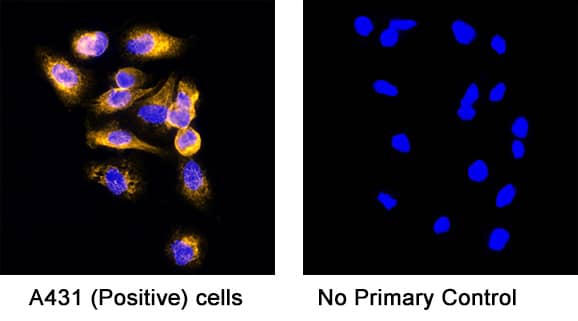
Host Cell Factor 1/HCFC1 in HeLa Human Cell Line. Host Cell Factor 1/HCFC1 was detected in immersion fixed HeLa human cervical epithelial carcinoma cell line using Goat Anti-Human Host Cell Factor 1/HCFC1 Antigen Affinity-purified Polyclonal Antibody (Catalog # AF6254) at 10 µg/mL for 3 hours at room temperature. Cells were stained using the NorthernLights™ 557-conjugated Anti-Goat IgG Secondary Antibody (red; Catalog # NL001) and counterstained with DAPI (blue). Specific staining was localized to nuclei. View our protocol for Fluorescent ICC Staining of Cells on Coverslips.
.") View Larger
View Larger
Host Cell Factor 1/HCFC1 in Human Colon Cancer Tissue. Host Cell Factor 1/HCFC1 was detected in immersion fixed paraffin-embedded sections of human colon cancer tissue using Goat Anti-Human Host Cell Factor 1/HCFC1 Antigen Affinity-purified Polyclonal Antibody (Catalog # AF6254) at 10 µg/mL overnight at 4 °C. Tissue was stained using the Anti-Goat HRP-DAB Cell & Tissue Staining Kit (brown; Catalog # CTS008) and counterstained with hematoxylin (blue). Specific staining was localized to nuclei of epithelial cells. View our protocol for Chromogenic IHC Staining of Paraffin-embedded Tissue Sections.
Preparation and Storage
- 12 months from date of receipt, -20 to -70 °C as supplied.
- 1 month, 2 to 8 °C under sterile conditions after reconstitution.
- 6 months, -20 to -70 °C under sterile conditions after reconstitution.
Background: Host Cell Factor 1/HCFC1
HCF-1 (Host cell factor; also called C1 factor and VCAF) is a blanket name for a group of polypeptides that are generated through the targeted proteolysis of a large 300 kDa transcriptional coactivator precursor. The precursor is widely expressed, and its products are posited to participate in multiple activities, such as mRNA processing, transcriptional coactivation, and cell cycle progression through G0/G1 transition. Human HCF-1 is 2035 amino acids (aa) in length. It is modular in structure, and contains a Kelch-repeat region (aa 32-313), an SP1/GABP basic binding sequence (aa 478-875), an acidic transactivation domain (aa 1530-1735), and a C-terminal NLS-containing Trp/Tyr/Phe-rich region (aa 1760-2035). Situated between the basic and acidic regions is an HCF repeat region (aa 1010-1439) that contains six 26 aa repeats that undergo autocatalytic cleavage. HCF-1 is O-glycosylated, phosphorylated and acetylated. In quiescent cells, the full-length 300 kDa precursor can be found in the cytoplasm, along with a presumed 50 kDa fragment that appears to represent some sequence between aa 300-1000. In activated cells, HCF-1 appears in the nucleus, and undergoes autocatalysis at one or more sites occurs distal to Glu at position 1019, 1081, 1110, 1295, 1323, and 1423. This generates multiple fragments that noncovalently associate with each other and appear as bands in SDS-Page that range from 100-175 kDa in size. This association is quite vigorous and requires aggressive treatment to achieve denaturation. There is one potential isoform that contains an alternative start site at Met100, a second isoform that shows a deletion of aa 1072-1101, a third isoform that exhibits a Leu substitution for aa 428-2035, and a final isoform that shows a deletion of aa 382-450. Over aa 1626-1836, human HCF-1 shares 95% aa identity with mouse HCF-1.
Product Datasheets
Citation for Human Host Cell Factor 1/HCFC1 Antibody
R&D Systems personnel manually curate a database that contains references using R&D Systems products. The data collected includes not only links to publications in PubMed, but also provides information about sample types, species, and experimental conditions.
1 Citation: Showing 1 - 1
-
Heat-Shock Protein 90 Controls the Expression of Cell-Cycle Genes by Stabilizing Metazoan-Specific Host-Cell Factor HCFC1
Authors: A Antonova, B Hummel, A Khavaran, DM Redhaber, F Aprile-Gar, P Rawat, K Gundel, M Schneck, EC Hansen, J Mitschke, G Mittler, C Miething, R Sawarkar
Cell Rep, 2019-11-05;29(6):1645-1659.e9.
Species: Human
Sample Types: Cell Lysates
Applications: Western Blot
FAQs
No product specific FAQs exist for this product, however you may
View all Antibody FAQsIsotype Controls
Reconstitution Buffers
Secondary Antibodies
Reviews for Human Host Cell Factor 1/HCFC1 Antibody
There are currently no reviews for this product. Be the first to review Human Host Cell Factor 1/HCFC1 Antibody and earn rewards!
Have you used Human Host Cell Factor 1/HCFC1 Antibody?
Submit a review and receive an Amazon gift card.
$25/€18/£15/$25CAN/¥75 Yuan/¥2500 Yen for a review with an image
$10/€7/£6/$10 CAD/¥70 Yuan/¥1110 Yen for a review without an image
