Human/Mouse/Rat Peroxiredoxin 1 Antibody Summary
Met1-Lys199
Accession # Q06830
*Small pack size (-SP) is supplied either lyophilized or as a 0.2 µm filtered solution in PBS.
Customers also Viewed


 Bioactivity")


Applications
Please Note: Optimal dilutions should be determined by each laboratory for each application. General Protocols are available in the Technical Information section on our website.
Scientific Data
 View Larger
Detection of Human/Mouse/Rat Peroxiredoxin 1 by Western Blot. Western blot shows lysates of Jurkat human acute T cell leukemia cell line, Raji human Burkitt's lymphoma cell line, MCF-7 human breast cancer cell line, CH-1 mouse B cell lymphoma cell line, A20 mouse B cell lymphoma cell line, and L6 rat myoblast cell line. PVDF membrane was probed with 0.2 µg/mL of Goat Anti-Human/Mouse/Rat Peroxiredoxin 1 Antigen Affinity-purified Polyclonal Antibody (Catalog # AF3488) followed by HRP-conjugated Anti-Goat IgG Secondary Antibody (HAF109). A specific band was detected for Peroxiredoxin 1 at approximately 22 kDa (as indicated). This experiment was conducted using Immunoblot Buffer Group 2.
 View Larger
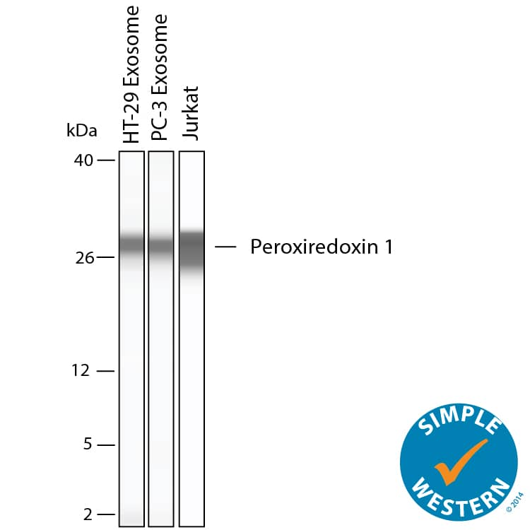
Detection of Human Peroxiredoxin 1 by Simple WesternTM. Simple Western shows lysates of Exosome Standards (HT‑29) (NBP3-11685), Exosome Standards (PC‑3) (NBP2-49856), and Jurkat human acute T cell leukemia cell line, loaded at 0.5 mg/ml. A specific band was detected for Peroxiredoxin 1 at approximately 28 kDa (as indicated) using 10 µg/mL of Goat Anti-Human/Mouse/Rat Peroxiredoxin 1 Antigen Affinity-purified Polyclonal Antibody (Catalog # AF3488). This experiment was conducted under reducing conditions and using the 2-40kDa separation system.
 View Larger
Western Blot Shows Human Peroxiredoxin 1 Specificity Using Knockout Cell Line. Western blot shows lysates of U2OS human osteosarcoma cell line and Peroxiredoxin 1 knockout U2OS cell line (KO). Nitrocellulose membrane was probed with 0.2 µg/mL of Goat Anti-Human/Mouse/Rat Peroxiredoxin 1 Antigen Affinity-purified Polyclonal Antibody (Catalog # AF3488) followed by HRP-conjugated Anti-Goat IgG Secondary Antibody. A specific band was detected for Peroxiredoxin 1 at approximately 22 kDa (as indicated) in the parental U2OS cell line, but is not detectable in knockout U2OS cell line. The Ponceau stained transfer of the blot is shown. This experiment was conducted under reducing conditions. Image, protocol, and testing courtesy of YCharOS Inc. See ycharos.com for additional details.
 View Larger
Detection of Human Peroxiredoxin 1 by Simple WesternTM. Simple Western lane view shows lysates of Jurkat human acute T cell leukemia cell line and Raji human Burkitt's lymphoma cell line, loaded at 0.2 mg/mL. A specific band was detected for Peroxiredoxin 1 at approximately 29 kDa (as indicated) using 2 µg/mL of Goat Anti-Human/Mouse/Rat Peroxiredoxin 1 Antigen Affinity-purified Polyclonal Antibody (Catalog # AF3488) followed by 1:50 dilution of HRP-conjugated Anti-Goat IgG Secondary Antibody (HAF109). This experiment was conducted under reducing conditions and using the 12-230 kDa separation system.
Preparation and Storage
- 12 months from date of receipt, -20 to -70 °C as supplied.
- 1 month, 2 to 8 °C under sterile conditions after reconstitution.
- 6 months, -20 to -70 °C under sterile conditions after reconstitution.
Background: Peroxiredoxin 1
Human Peroxiredoxin 1 (Prx-1 or PRDX1; also Thioredoxin Peroxidase 2) is a 22 kDa antioxidant enzyme that belongs to the typical 2-Cys class of the THP/ahpC family of proteins. The molecule is 199 amino acids (aa) in length, and has two catalytic cysteines, one at Cys52, and a second at Cys173. Prx-1 is an obligate homodimer. Inactive, it is apparently noncovalently associated. Upon peroxide binding to Cys52 of subunit 1, the Cys173 of subunit 2 interacts with Cys52 of subunit 1 to complete the antioxidation, generating a disulfide bond between Cys52 and Cys173. Subsequent reduction restores the subunits to the basal state. There are apparently two additional isoforms. One shows a premature truncation after aa 171, while the second shows a deletion of aa 21 - 121. Human Prx-1 shows 96% and 98% amino acid identity to mouse and rat Prx-1, respectively.
Product Datasheets
Citations for Human/Mouse/Rat Peroxiredoxin 1 Antibody
R&D Systems personnel manually curate a database that contains references using R&D Systems products. The data collected includes not only links to publications in PubMed, but also provides information about sample types, species, and experimental conditions.
2
Citations: Showing 1 - 2
Filter your results:
Filter by:
-
Electrophiles modulate glutathione reductase activity via alkylation and upregulation of glutathione biosynthesis
Authors: S Jobbagy, DA Vitturi, SR Salvatore, L Turell, MF Pires, E Kansanen, C Batthyany, JR Lancaster, BA Freeman, FJ Schopfer
Redox Biol, 2018-11-22;21(0):101050.
Species: Mouse
Sample Types: Cell Lysates
Applications: Western Blot -
The impact of tyrosine kinase 2 (Tyk2) on the proteome of murine macrophages and their response to lipopolysaccharide (LPS).
Authors: Radwan M, Miller I, Grunert T, Marchetti-Deschmann M, Vogl C, O'Donoghue N, Dunn MJ, Kolbe T, Allmaier G, Gemeiner M, Muller M, Strobl B
Proteomics, 2008-09-01;8(17):3469-85.
Species: Mouse
Sample Types: Cell Lysates
Applications: Western Blot
FAQs
No product specific FAQs exist for this product, however you may
View all Antibody FAQsIsotype Controls
Reconstitution Buffers
Secondary Antibodies
Reviews for Human/Mouse/Rat Peroxiredoxin 1 Antibody
There are currently no reviews for this product. Be the first to review Human/Mouse/Rat Peroxiredoxin 1 Antibody and earn rewards!
Have you used Human/Mouse/Rat Peroxiredoxin 1 Antibody?
Submit a review and receive an Amazon gift card.
$25/€18/£15/$25CAN/¥75 Yuan/¥2500 Yen for a review with an image
$10/€7/£6/$10 CAD/¥70 Yuan/¥1110 Yen for a review without an image

