Th17 Cell Markers
Click on one of the helper T cell subsets shown in the buttons below to see the markers that are most commonly used to identify that cell type.

Recommended Products
Overview
T helper type 17 (Th17) cells are a subset of CD4+ effector T cells that is required for host defense against specific fungi and extracellular bacteria. In mice, naïve CD4+ T cells differentiate into Th17 cells in the presence of IL-6 and TGF-beta, and IL-21 and IL-23 are required to stabilize the Th17 phenotype. In contrast, Th17 differentiation in humans requires IL-1 beta, IL-6, IL-21, and IL-23, but is less dependent on TGF-beta. Both mice and human Th17 cells are commonly identified as CD3+CD4+CD8- T cells that express TGF-beta R, IL-6 R alpha, IL-21 R, IL-23R, and the transcription factors, STAT3 and ROR-gamma t, the latter of which is the master transcriptional regulator required for Th17 cell development. In addition to these cell surface and intracellular markers, mouse and human Th17 cells can be distinguished from other CD4+ T cell subsets based on the production of IL-17A, IL-17F, IL-21, and IL-22. Human Th17 cells also secrete IL-26, a cytokine lacking a murine homologue. Cytokines secreted by Th17 cells induce the expression of secondary pro-inflammatory mediators and anti-microbial peptides. While Th17 cells play an important role in the elimination of harmful pathogens, unregulated expression of Th17-secreted cytokines has been linked to pathogenesis of autoimmune diseases such as inflammatory bowel disease, multiple sclerosis, psoriasis, and rheumatoid arthritis.
Data Examples
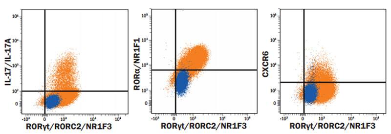
Detection of Cell Surface and Intracellular Markers on Human Th17 Cells by Flow Cytometry. CD4+ T cells were isolated from total human peripheral blood mononuclear cells using a cell selection protocol, such as the one found in the MagCellect™ Human CD4+ T Cell Isolation Kit (R&D Systems, Catalog # MAGH102). Isolated cells were incubated at 37 ˚C for seven days in media containing Recombinant Human IL-2 (R&D Systems, Catalog # 202-IL), Human TGF-beta 1 (R&D Systems, Catalog # 100-B), Recombinant Human IL-23 (R&D Systems, Catalog # 1290-IL), Recombinant Human IL-6 (R&D Systems, Catalog # 206-IL), Recombinant Human IL-1 beta (R&D Systems, Catalog # 201-LB), and Anti-Human IFN-gamma Antibody (R&D Systems, Catalog # AF-285-NA), followed by stimulation with 50 ng/mL PMA, 200 ng/mL calcium ionomycin, and 3 µM monensin for several hours. Live, single, CD4+ cells are shown in the dot plots, determined using a fixable viability dye, doublet exclusion, and staining with an Alexa Fluor® 594-conjugated Mouse Anti-Human CD4 Monoclonal Antibody (R&D Systems, Catalog # FAB3791T). The cells were additionally stained using an Alexa Fluor® 700-conjugated Mouse Anti-Human IL-17/IL-17A Monoclonal Antibody (R&D Systems, Catalog # IC3171N), a PE-conjugated Mouse Anti-Human ROR alpha/NR1F1 Monoclonal Antibody (R&D Systems, Catalog # IC8924P), an Alexa Fluor® 488-conjugated Rabbit Anti-Human/Mouse ROR gamma t Monoclonal Antibody (R&D Systems, Catalog # IC9125G), and an APC-conjugated Mouse Anti-Human CXCR6 Monoclonal Antibody (R&D Systems, Catalog # FAB699A). Dot plots show the relative IL-17A+, ROR alpha+, CXCR6+, and ROR gamma t+ cells in CD4+ resting (blue dots, lower left) and Th17-differentiated (orange dots, upper right quadrants) cell populations. Quadrant markers were set based on staining with the appropriate isotype controls (R&D Systems, Catalog # IC003T, # IC0041N, # IC0041P, # IC1051G, and # IC0041A). Note: The five fluorochrome-conjugated antibodies and buffers used for cell surface and intracellular staining of human Th17 cells using this protocol are also provided together in the FlowX™ Human Th17 Cell Multi-Color Flow Cytometry Kit (R&D Systems, Catalog # FMC007B).

