Human IGFBP-4 Antibody Summary
Asp22-Glu258
Accession # AAA62670
Customers also Viewed
Applications
Please Note: Optimal dilutions should be determined by each laboratory for each application. General Protocols are available in the Technical Information section on our website.
Scientific Data
 View Larger
IGFBP‑4 in Human Placenta. IGFBP-4 was detected in immersion fixed paraffin-embedded sections of human placenta (chorionic villi) using 5 µg/mL Goat Anti-Human IGFBP-4 Antigen Affinity-purified Polyclonal Antibody (Catalog # AF804) overnight at 4 °C. Before incubation with the primary antibody tissue was subjected to heat-induced epitope retrieval using Antigen Retrieval Reagent-Basic (Catalog # CTS013). Tissue was stained with the Anti-Goat HRP-AEC Cell & Tissue Staining Kit (red; Catalog # CTS009) and counterstained with hematoxylin (blue). View our protocol for Chromogenic IHC Staining of Paraffin-embedded Tissue Sections.
 View Larger
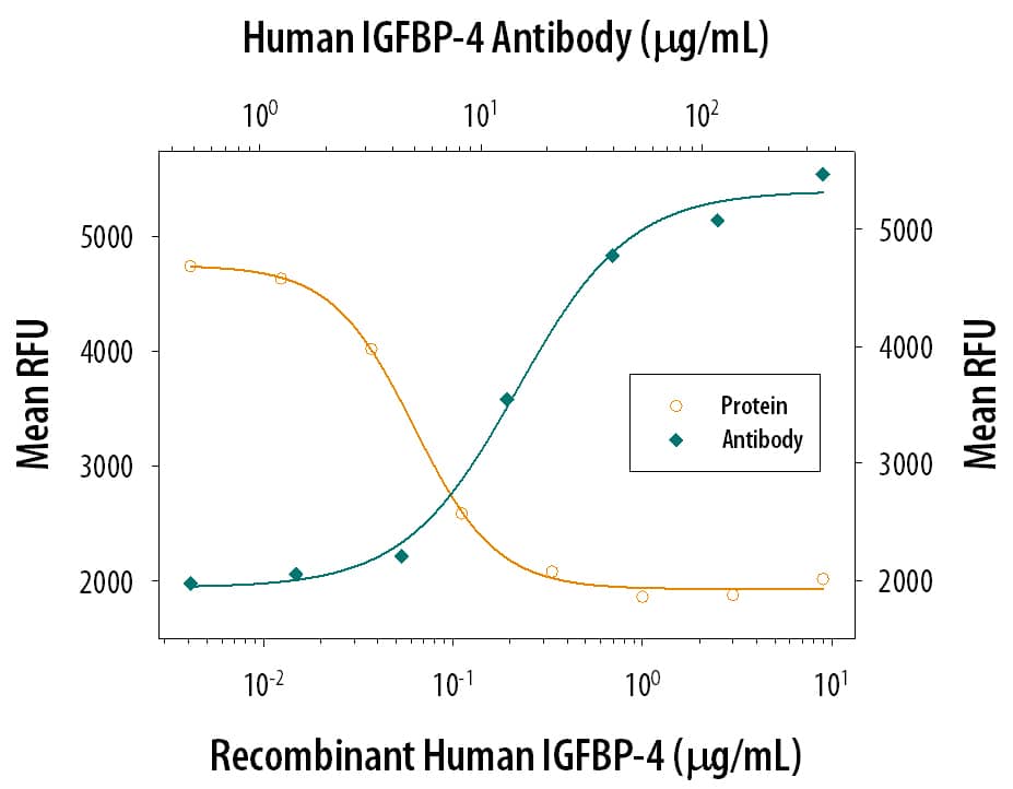
IGFBP‑4 Inhibition of IGF‑II-dependent Cell Proliferation and Neutralization by Human IGFBP‑4 Antibody. Recombinant Human IGFBP-4 (Catalog # 804-GB) inhibits Recombinant Human IGF-II (Catalog # 292-G2) induced proliferation in the MCF-7 human breast cancer cell line in a dose-dependent manner (orange line). Inhibition of Recombinant Human IGF-II (14 ng/mL) activity elicited by Recombinant Human IGFBP-4 (0.3 µg/mL) is neutralized (green line) by increasing concentrations of Goat Anti-Human IGFBP-4 Antigen Affinity-purified Polyclonal Antibody (Catalog # AF804). The ND50 is typically 12-60 µg/mL.
 View Larger
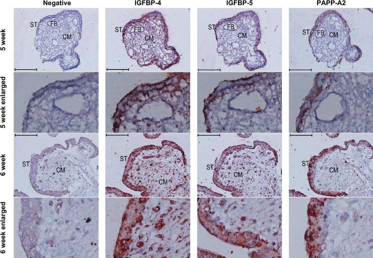
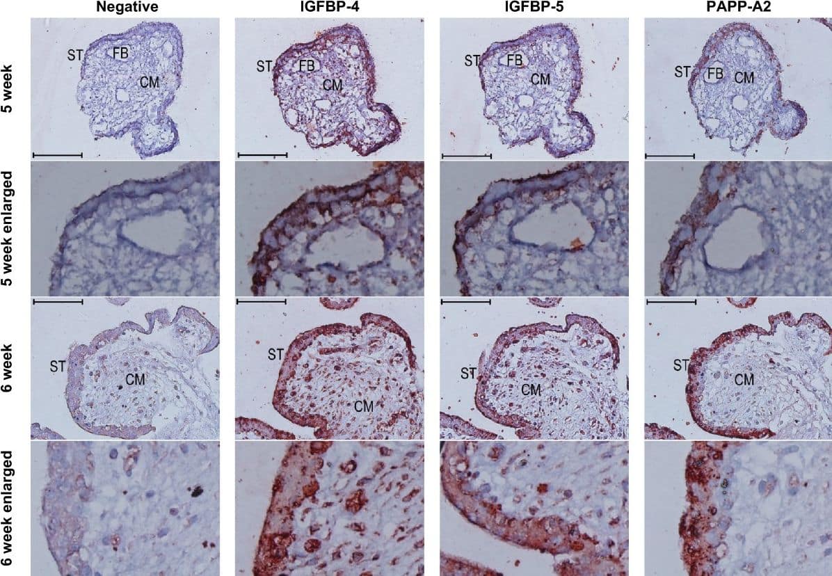
Detection of Human IGFBP-4 by Immunohistochemistry Immunoreactivity (red colour) against IGFBP-4, −5, and PAPPA2 in the syncytiotrophoblast of first trimester placental villi. Serial cross sections of placental villi at 5 and 6 weeks of gestation (7 and 13 weeks not shown) were stained for IGFBP-4, IGFBP-5, and PAPP-A2. Negative controls used non-specific goat IgG in the place of the primary antibodies. FB = fetal blood vessel, CM = chorionic mesoderm, ST = syncytiotrophoblast. Scale bars denote 100 μm. Image collected and cropped by CiteAb from the following open publication (https://pubmed.ncbi.nlm.nih.gov/25475528), licensed under a CC-BY license. Not internally tested by R&D Systems.
Preparation and Storage
- 12 months from date of receipt, -20 to -70 °C as supplied.
- 1 month, 2 to 8 °C under sterile conditions after reconstitution.
- 6 months, -20 to -70 °C under sterile conditions after reconstitution.
Background: IGFBP-4
Human IGF binding protein 4 (IGFBP-4) was isolated from human plasma based on its ability to bind immobilized IGF-I. Human IGFBP-4 cDNA encodes a 258 amino acid (aa) residue precursor protein with a predicted 21 aa residue signal peptide that is processed to generate the 237 aa residue mature human IGFBP-4. The human IGFBP-4 contains a potential N-linked glycosylation site and shares approximately 90% amino acid sequence identity with both the mouse and rat IGFBP-4.
According to the nomenclature of IGFBPs defined at the 4th International Symposium of IGFs (1997, Tokyo), six high-affinity IGF binding proteins
(IGFBP-1, -2, -3, ‑4, ‑5, ‑6), and four IGFBP-related proteins (IGFBPr-1, - 2, -3, -4) have been identified. All IGFBPs have a high cysteine content and share conserved cysteine residues that are clustered in the amino- and carboxy-terminal third of the molecule. IGFBPs have been shown to either inhibit or enhance the biological activities of IGF, or act in an IGF-independent manner. Post-translational modification of IGFBPs, including phosphorylation and proteolysis, have been shown to modify the affinities of the binding proteins for IGF and may indirectly regulate IGF actions.
- Jones, J. I. and D.R. Clemons (1995) Endocrine Rev. 16:3.
- Kelly, K.M. et al. (1996) Intl. J. Biochem. Cell Biol. 28:619.
- Kim, H-S. et al. (1997) Proc. Natl. Acad. Sci. USA 94:12981.
Product Datasheets
Citations for Human IGFBP-4 Antibody
R&D Systems personnel manually curate a database that contains references using R&D Systems products. The data collected includes not only links to publications in PubMed, but also provides information about sample types, species, and experimental conditions.
4
Citations: Showing 1 - 4
Filter your results:
Filter by:
-
EWS-FLI-1 creates a cell surface microenvironment conducive to IGF signaling by inducing pappalysin-1
Authors: P Jayabal, PJ Houghton, Y Shiio
Genes Cancer, 2017-11-01;8(11):762-770.
Species: Human
Sample Types: Cell Lysates
Applications: Western Blot -
IGFBP-4 and -5 are expressed in first-trimester villi and differentially regulate the migration of HTR-8/SVneo cells.
Authors: Crosley, Erin J, Dunk, Caroline, Beristain, Alexande, Christians, Julian K
Reprod Biol Endocrinol, 2014-12-04;12(0):123.
Species: Human
Sample Types: Whole Tissue
Applications: IHC-P -
Fetal baboon sex-specific outcomes in adipocyte differentiation at 0.9 gestation in response to moderate maternal nutrient reduction.
Authors: Tchoukalova Y, Krishnapuram R, White U, Burk D, Fang X, Nijland M, Nathanielsz P
Int J Obes (Lond), 2013-06-10;38(2):224-30.
Species: Primate - Papio cynocephalus (Yellow Baboon)
Sample Types: Whole Cells
Applications: Western Blot -
The role of matrix metalloproteinase-7 in redefining the gastric microenvironment in response to Helicobacter pylori.
Authors: McCaig C, Duval C, Hemers E, Steele I, Pritchard DM, Przemeck S, Dimaline R, Ahmed S, Bodger K, Kerrigan DD, Wang TC, Dockray GJ, Varro A
Gastroenterology, 2006-03-06;130(6):1754-63.
Species: Human
Sample Types: Cell Lysates
Applications: Western Blot
FAQs
No product specific FAQs exist for this product, however you may
View all Antibody FAQsIsotype Controls
Reconstitution Buffers
Secondary Antibodies
Reviews for Human IGFBP-4 Antibody
Average Rating: 3 (Based on 1 Review)
Have you used Human IGFBP-4 Antibody?
Submit a review and receive an Amazon gift card.
$25/€18/£15/$25CAN/¥75 Yuan/¥2500 Yen for a review with an image
$10/€7/£6/$10 CAD/¥70 Yuan/¥1110 Yen for a review without an image
Filter by:


