Mouse Sca-1/Ly6 Antibody Summary
Leu27-Gly119
Accession # CAA28351
Customers also Viewed
Applications
Please Note: Optimal dilutions should be determined by each laboratory for each application. General Protocols are available in the Technical Information section on our website.
Scientific Data
 View Larger
Sca‑1/Ly6 in Mouse Splenocytes. Sca-1/Ly6 was detected in immersion fixed mouse splenocytes using 10 µg/mL Goat Anti-Mouse Sca-1/Ly6 Antigen Affinity-purified Polyclonal Antibody (Catalog # AF1226) for 3 hours at room temperature. Cells were stained with the NorthernLights™ 557-conjugated Anti-Goat IgG Secondary Antibody (red; Catalog # NL001) and counterstained with DAPI (blue). View our protocol for Fluorescent ICC Staining of Non-adherent Cells.
 View Larger
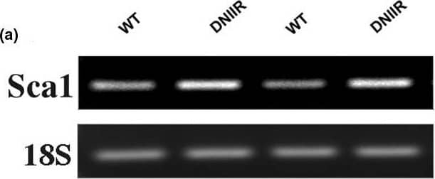
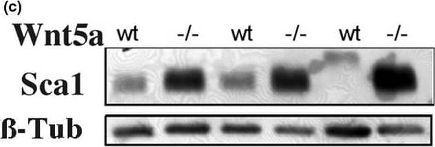
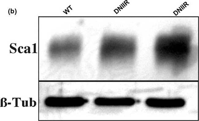
Detection of Mouse Sca-1/Ly6 by Western Blot Sca1, a marker of mammary stem/progenitor cells, is increased in DNIIR and Wnt5a-/- glands. mRNA and protein were extracted from several wild type (WT) and MT-DNIIR (DNIIR) mice. (a) Sca1 mRNA levels were determined by semi-quantitative RT-PCR and (b) protein levels were determined by western blot. Sca1 levels were increased in DNIIR glands relative to controls. (c) Sca1 protein levels were determined in WT and Wnt5a null (-/-) glands using western blot analysis. Sca1 levels were increased in Wnt5a-null tissue relative to controls. 18S and beta -Tubulin ( beta -Tub) were used as normalisation controls. DNIIR = dominant-negative transforming growth factor-beta type II receptor; RT-PCR = reverse transcripase polymerase chain reaction; Wnt = Wingless-related protein family. Image collected and cropped by CiteAb from the following open publication (https://pubmed.ncbi.nlm.nih.gov/19344510), licensed under a CC-BY license. Not internally tested by R&D Systems.
 View Larger
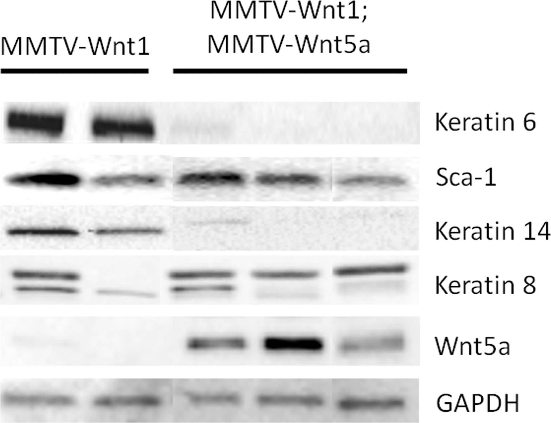
Detection of Mouse Sca-1/Ly6 by Western Blot Ectopic expression of Wnt5a results in low expression of K6 and K14.Expression of molecular markers for basal and luminal progenitors in MMTV-Wnt1 and MMTV-Wnt1;MMTV-Wnt5a mammary glands was compared by western blot. Glyceraldehyde 3-phosphate dehydrogenase (GAPDH) was used as a loading control. Each lane contains protein isolated from a separate mouse. Image collected and cropped by CiteAb from the following open publication (https://dx.plos.org/10.1371/journal.pone.0113247), licensed under a CC-BY license. Not internally tested by R&D Systems.
 View Larger
Detection of Mouse Sca-1/Ly6 by Western Blot Sca1, a marker of mammary stem/progenitor cells, is increased in DNIIR and Wnt5a-/- glands. mRNA and protein were extracted from several wild type (WT) and MT-DNIIR (DNIIR) mice. (a) Sca1 mRNA levels were determined by semi-quantitative RT-PCR and (b) protein levels were determined by western blot. Sca1 levels were increased in DNIIR glands relative to controls. (c) Sca1 protein levels were determined in WT and Wnt5a null (-/-) glands using western blot analysis. Sca1 levels were increased in Wnt5a-null tissue relative to controls. 18S and beta -Tubulin ( beta -Tub) were used as normalisation controls. DNIIR = dominant-negative transforming growth factor-beta type II receptor; RT-PCR = reverse transcripase polymerase chain reaction; Wnt = Wingless-related protein family. Image collected and cropped by CiteAb from the following open publication (https://pubmed.ncbi.nlm.nih.gov/19344510), licensed under a CC-BY license. Not internally tested by R&D Systems.
 View Larger
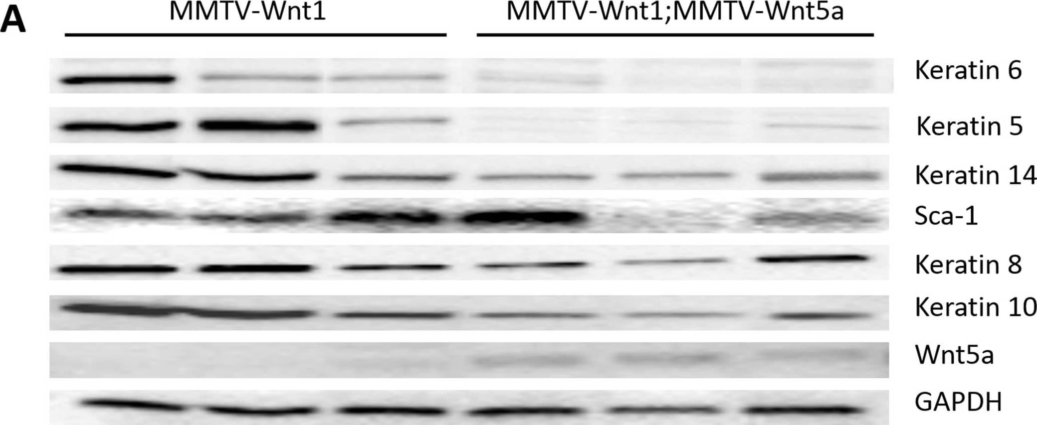
Detection of Mouse Sca-1/Ly6 by Western Blot Wnt5a expressing tumors demonstrate a decrease in markers of the basal tumor subtype.(A) Western blot using protein lysates isolated from the epithelium of MMTV-Wnt1 and MMTV-1;MMTV-Wnt5a mammary tumors. The expression of molecular markers of basal and luminal tumor subtypes were compared. Keratin 6 and Keratin 5 were strongly down-regulated in Wnt5a expressing tumors. Glyceraldehyde 3-phosphate dehydrogenase (GAPDH) was used as a loading control. (B–D) Immunostaining for K6. Sections from MMTV-Wnt1 (B) and MMTV-1;MMTV-Wnt5a (C) tumors were stained with anti-Keratin 6 antibody using immunofluorescence (K6 = green; nuclei = blue). The percentage of cells expressing K6 was determined and graphed (D). Values are means +/− standard error (n = 6 MMTV-Wnt1, 3 fields per tumor; n = 5 MMTV-Wnt1;MMTV-Wnt5a, 3 fields per tumor). MMTV-Wnt1;MMTV-Wnt5a tumors demonstrated a significant decrease in K6-expressing cells as measured by T-test (* = p<0.05). Image collected and cropped by CiteAb from the following open publication (https://dx.plos.org/10.1371/journal.pone.0113247), licensed under a CC-BY license. Not internally tested by R&D Systems.
 View Larger
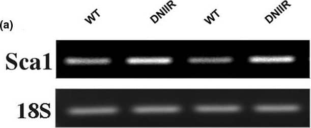
Detection of Mouse Sca-1/Ly6 by Western Blot Sca1, a marker of mammary stem/progenitor cells, is increased in DNIIR and Wnt5a-/- glands. mRNA and protein were extracted from several wild type (WT) and MT-DNIIR (DNIIR) mice. (a) Sca1 mRNA levels were determined by semi-quantitative RT-PCR and (b) protein levels were determined by western blot. Sca1 levels were increased in DNIIR glands relative to controls. (c) Sca1 protein levels were determined in WT and Wnt5a null (-/-) glands using western blot analysis. Sca1 levels were increased in Wnt5a-null tissue relative to controls. 18S and beta -Tubulin ( beta -Tub) were used as normalisation controls. DNIIR = dominant-negative transforming growth factor-beta type II receptor; RT-PCR = reverse transcripase polymerase chain reaction; Wnt = Wingless-related protein family. Image collected and cropped by CiteAb from the following open publication (https://pubmed.ncbi.nlm.nih.gov/19344510), licensed under a CC-BY license. Not internally tested by R&D Systems.
Preparation and Storage
- 12 months from date of receipt, -20 to -70 °C as supplied.
- 1 month, 2 to 8 °C under sterile conditions after reconstitution.
- 6 months, -20 to -70 °C under sterile conditions after reconstitution.
Background: Sca-1/Ly6
This antibody reacts with Sca-1, an 18 kDa phosphatidylinositol-anchored protein that is a member of the lymphocyte antigen 6 (Ly-6) family (1). Sca-1 is encoded by the strain-specific Ly-6 E/A allelic gene. Its expression on multipotent hematopoietic stem cells (HSC) has been used as a marker of HSC in mice of both Ly‑6 haplotypes (2, 3). This antibody is frequently used in combination with lineage depletion antibodies to identify and isolate murine HSC. Sca-1-positive HSC can be found in the adult bone marrow, fetal liver and mobilized peripheral blood and spleen in the adult animal (2‑7). However, Sca-1 has also been discovered in several non‑hematopoietic tissues (1) and can be used to enrich progenitor cell populations other than HSC (8). It is suggested that Sca-1 could be involved in regulating both B and T cell activation (9‑12).
- Van de Rijn, M. et al. (1989) Proc. Natl.Acad. Sci. USA 86:4634.
- Spangrude, G.I. et al. (1988) Science 241:58.
- Spangrude, G.I. et al. (1993) Blood 82:3327.
- Morrison, S.J. et al. (1995) Proc. Natl. Acad. Sci. USA 92:10302.
- Kawamoto, H. et al. (1997) Int. Immunol. 9:1011.
- Yamamoto, Y. et al. (1996) Blood 88:445.
- Morrison, S.J. et al. (1997) Proc. Natl. Acad. Sci. USA 94:1908.
- Welm, B.E. et al. (2002) Dev. Biol. 245:42.
- Codias, E.K. et al. (1990) J. Immunol. 144:2197.
- Malek, T.R. et al. (1986) J. Exp. Med. 164:709 .
- Codias, E.K. et al. (1990) J. Immunol. 145:1407.
- Flood, P.M. et al. (1990) J. Exp. Med. 172:115.
Product Datasheets
Citations for Mouse Sca-1/Ly6 Antibody
R&D Systems personnel manually curate a database that contains references using R&D Systems products. The data collected includes not only links to publications in PubMed, but also provides information about sample types, species, and experimental conditions.
29
Citations: Showing 1 - 10
Filter your results:
Filter by:
-
Smart Hydrogels for the Augmentation of Bone Regeneration by Endogenous Mesenchymal Progenitor Cell Recruitment
Authors: Philipp S. Lienemann, Queralt Vallmajo‐Martin, Panagiota Papageorgiou, Ulrich Blache, Stéphanie Metzger, Anna‐Sofia Kiveliö et al.
Advanced Science
-
Polyclonal regeneration of mouse bone marrow endothelial cells after irradiative conditioning
Authors: Skulimowska, I;Morys, J;Sosniak, J;Gonka, M;Gulati, G;Sinha, R;Kowalski, K;Mosiolek, S;Weissman, IL;Jozkowicz, A;Szade, A;Szade, K;
Cell reports
Species: Mouse, Transgenic Mouse
Sample Types: Whole Tissue
Applications: Immunohistochemistry -
Senescent fibroblasts in the tumor stroma rewire lung cancer metabolism and plasticity
Authors: Lee, JY;Reyes, N;Woo, SH;Goel, S;Stratton, F;Kuang, C;Mansfield, AS;LaFave, LM;Peng, T;
bioRxiv : the preprint server for biology
Species: Transgenic Mouse
Sample Types: Whole Tissue
Applications: Immunohistochemistry -
Bone marrow and periosteal skeletal stem/progenitor cells make distinct contributions to bone maintenance and repair
Authors: EC Jeffery, TLA Mann, JA Pool, Z Zhao, SJ Morrison
Cell Stem Cell, 2022-10-21;29(11):1547-1561.e6.
Species: Mouse
Sample Types: Whole Tissue
Applications: IHC -
CXCL12/CXCR4-Rac1-mediated migration of osteogenic precursor cells contributes to pathological new bone formation in ankylosing spondylitis
Authors: H Cui, Z Li, S Chen, X Li, D Chen, J Wang, Z Li, W Hao, F Zhong, K Zhang, Z Zheng, Z Zhan, H Liu
Science Advances, 2022-04-06;8(14):eabl8054.
Species: Mouse, Transgenic Mouse
Sample Types: Whole Tissue
Applications: IHC -
Bone marrow-independent adventitial macrophage progenitor cells contribute to angiogenesis
Authors: F Kleefeldt, B Upcin, H Bömmel, C Schulz, G Eckner, J Allmanritt, J Bauer, B Braunger, U Rueckschlo, S Ergün
Cell Death & Disease, 2022-03-09;13(3):220.
Species: Mouse
Sample Types: Whole Cells
Applications: ICC -
Fecal microbiota transplantation ameliorates atherosclerosis in mice with C1q/TNF-related protein 9 genetic deficiency
Authors: ES Kim, BH Yoon, SM Lee, M Choi, EH Kim, BW Lee, SY Kim, CG Pack, YH Sung, IJ Baek, CH Jung, TB Kim, JY Jeong, CH Ha
Experimental & Molecular Medicine, 2022-02-03;0(0):.
Species: Mouse
Sample Types: Whole Tissue
Applications: IHC -
Distinct Expression Patterns of Cxcl12 in Mesenchymal Stem Cell Niches of Intact and Injured Rodent Teeth
Authors: P Pagella, C Nombela-Ar, TA Mitsiadis
International Journal of Molecular Sciences, 2021-03-16;22(6):.
Species: Mouse
Sample Types: Whole Tissue
Applications: IHC -
Multicolor quantitative confocal imaging cytometry
Authors: DL Coutu, KD Kokkaliari, L Kunz, T Schroeder
Nat. Methods, 2017-11-13;15(1):39-46.
Species: Mouse
Sample Types: Whole Tissue
Applications: IHC -
Bone marrow-derived cells and their conditioned medium induce microvascular repair in uremic rats by stimulation of endogenous repair mechanisms
Authors: L Golle, HU Gerth, K Beul, B Heitplatz, P Barth, M Fobker, H Pavenstädt, GS Di Marco, M Brand
Sci Rep, 2017-08-25;7(1):9444.
Species: Rat
Sample Types: Whole Cells
Applications: Flow Cytometry -
Dipeptidyl Peptidase-4 Regulates Hematopoietic Stem Cell Activation in Response to Chronic Stress
Authors: E Zhu, L Hu, H Wu, L Piao, G Zhao, A Inoue, W Kim, C Yu, W Xu, YK Bando, X Li, Y Lei, CN Hao, K Takeshita, WS Kim, K Okumura, T Murohara, M Kuzuya, XW Cheng
J Am Heart Assoc, 2017-07-14;6(7):.
Species: Mouse
Sample Types: Whole Cells
Applications: ICC -
Systemic application of sirolimus prevents neointima formation not via a direct anti-proliferative effect but via its anti-inflammatory properties
Authors: JM Daniel, J Dutzmann, H Brunsch, J Bauersachs, R Braun-Dull, DG Sedding
Int. J. Cardiol., 2017-03-14;0(0):.
Species: Mouse
Sample Types: Whole Tissue
Applications: IHC -
Pluripotent stem cells derived from mouse and human white mature adipocytes.
Authors: Jumabay M, Abdmaulen R, Ly A, Cubberly M, Shahmirian L, Heydarkhan-Hagvall S, Dumesic D, Yao Y, Bostrom K
Stem Cells Transl Med, 2014-01-06;3(2):161-71.
Species: Mouse
Sample Types: Whole Cells
Applications: ICC -
Distinct fibroblast lineages determine dermal architecture in skin development and repair.
Authors: Driskell R, Lichtenberger B, Hoste E, Kretzschmar K, Simons B, Charalambous M, Ferron S, Herault Y, Pavlovic G, Ferguson-Smith A, Watt F
Nature, 2013-12-12;504(7479):277-81.
Species: Mouse
Sample Types: Whole Tissue
Applications: IHC-Fr -
Age dependence of lung mesenchymal stromal cell dynamics following pneumonectomy.
Authors: Paxson J, Gruntman A, Davis A, Parkin C, Ingenito E, Hoffman A
Stem Cells Dev, 2013-08-30;22(24):3214-25.
Species: Mouse
Sample Types: Whole Tissue
Applications: IHC-P -
Structural basis of digoxin that antagonizes RORgamma t receptor activity and suppresses Th17 cell differentiation and interleukin (IL)-17 production.
Authors: Fujita-Sato S, Ito S, Isobe T, Ohyama T, Wakabayashi K, Morishita K, Ando O, Isono F
J. Biol. Chem., 2011-07-06;286(36):31409-17.
Species: Mouse
Sample Types: Whole Tissue
Applications: IHC-Fr -
Stromal cell-derived factor-1 enhances distraction osteogenesis-mediated skeletal tissue regeneration through the recruitment of endothelial precursors.
Authors: Fujio M, Yamamoto A, Ando Y, Shohara R, Kinoshita K, Kaneko T, Hibi H, Ueda M
Bone, 2011-06-29;49(4):693-700.
Species: Mouse
Sample Types: Whole Tissue
Applications: IHC-Fr -
Three-dimensional culture of mouse renal carcinoma cells in agarose macrobeads selects for a subpopulation of cells with cancer stem cell or cancer progenitor properties.
Authors: Smith BH, Gazda LS, Conn BL
Cancer Res., 2011-01-24;71(3):716-24.
Species: Mouse
Sample Types: Whole Tissue
Applications: IHC -
Prominin-1/CD133+ lung epithelial progenitors protect from bleomycin-induced pulmonary fibrosis.
Authors: Germano D, Blyszczuk P, Valaperti A, Kania G, Dirnhofer S, Landmesser U, Luscher TF, Hunziker L, Zulewski H, Eriksson U
Am. J. Respir. Crit. Care Med., 2009-02-20;179(10):939-49.
Species: Mouse
Sample Types: Whole Cells
Applications: Flow Cytometry -
A Gata6-Wnt pathway required for epithelial stem cell development and airway regeneration.
Authors: Zhang Y, Goss AM, Cohen ED, Kadzik R, Lepore JJ, Muthukumaraswamy K, Yang J, DeMayo FJ, Whitsett JA, Parmacek MS, Morrisey EE
Nat. Genet., 2008-06-08;40(7):862-70.
Species: Mouse
Sample Types: Whole Cells
Applications: ICC -
Intestinal epithelial cell up-regulation of LY6 molecules during colitis results in enhanced chemokine secretion.
Authors: Flanagan K, Modrusan Z, Cornelius J, Chavali A, Kasman I, Komuves L, Mo L, Diehl L
J. Immunol., 2008-03-15;180(6):3874-81.
Species: Mouse
Sample Types: Whole Tissue
Applications: IHC-Fr -
The recruitment of primitive Lin(-) Sca-1(+), CD34(+), c-kit(+) and CD271(+) cells during the early intraperitoneal foreign body reaction.
Authors: Vranken I, De Visscher G, Lebacq A, Verbeken E, Flameng W
Biomaterials, 2007-11-26;29(7):797-808.
Species: Rat
Sample Types: Whole Cells
Applications: ICC -
Identification of pulmonary Oct-4+ stem/progenitor cells and demonstration of their susceptibility to SARS coronavirus (SARS-CoV) infection in vitro.
Authors: Ling TY, Kuo MD, Li CL, Yu AL, Huang YH, Wu TJ, Lin YC, Chen SH, Yu J
Proc. Natl. Acad. Sci. U.S.A., 2006-06-13;103(25):9530-5.
Species: Mouse
Sample Types: Whole Cells
Applications: ICC -
Identification of bronchioalveolar stem cells in normal lung and lung cancer.
Authors: Kim CF, Jackson EL, Woolfenden AE, Lawrence S, Babar I, Vogel S, Crowley D, Bronson RT, Jacks T
Cell, 2005-06-17;121(6):823-35.
Species: Mouse
Sample Types: Whole Cells
Applications: ICC -
Epidermal beta -catenin activation remodels the dermis via paracrine signalling to distinct fibroblast lineages
Authors: Beate M. Lichtenberger, Maria Mastrogiannaki, Fiona M. Watt
Nature Communications
-
Genetic lineage tracing of targeted cell populations during enthesis healing
Authors: Helen L. Moser, Anton Ph. Doe, Kristen Meier, Simon Garnier, Damien Laudier, Haruhiko Akiyama et al.
Journal of Orthopaedic Research®
-
Wnt5a Suppresses Tumor Formation and Redirects Tumor Phenotype in MMTV-Wnt1 Tumors
Authors: Stephanie L. Easter, Elizabeth H. Mitchell, Sarah E. Baxley, Renee Desmond, Andra R. Frost, Rosa Serra
PLoS ONE
-
Tgfbeta signaling is critical for maintenance of the tendon cell fate
Authors: Tan GK, Pryce BA, Stabio A et al.
Elife
-
Bone marrow-derived cells rescue salivary gland function in mice with head and neck irradiation
Authors: Yoshinori Sumita, Younan Liu, Saeed Khalili, Ola M. Maria, Dengsheng Xia, Sharon Key et al.
The International Journal of Biochemistry & Cell Biology
FAQs
No product specific FAQs exist for this product, however you may
View all Antibody FAQsIsotype Controls
Reconstitution Buffers
Secondary Antibodies
Reviews for Mouse Sca-1/Ly6 Antibody
Average Rating: 5 (Based on 1 Review)
Have you used Mouse Sca-1/Ly6 Antibody?
Submit a review and receive an Amazon gift card.
$25/€18/£15/$25CAN/¥75 Yuan/¥2500 Yen for a review with an image
$10/€7/£6/$10 CAD/¥70 Yuan/¥1110 Yen for a review without an image
Filter by:


