GFP Antibody Summary
aa 2-238
Customers also Viewed
Applications
Please Note: Optimal dilutions should be determined by each laboratory for each application. General Protocols are available in the Technical Information section on our website.
Scientific Data
 View Larger
Detection of GFP by Western Blot. Western blot shows synthetic eGFP, recombinant RANGAP-1 tagged with eYFP, recombinant GRPuv, and crystal jelly GFP. PVDF membrane was probed with 2 µg/mL of Mouse Anti-GFP Monoclonal Antibody (Catalog # MAB42402) followed by HRP-conjugated Anti-Mouse IgG Secondary Antibody (HAF018). A specific band was detected for GFP at approximately 28 and recombinant RANGAP-1 tagged with eYFP at 45 kDa (as indicated). This experiment was conducted under reducing conditions and using Western Blot Buffer Group 1.
 View Larger
GFP in HEK293 Human Cell Line Transfected with GFP. GFP was detected in immersion fixed HEK293 human embryonic kidney cell line transfected with GFP (green; right panel) using Mouse Anti-GFP Monoclonal Antibody (Catalog # MAB42402) at 8 µg/mL for 3 hours at room temperature. Cells were stained using the NorthernLights™ 557-conjugated Anti-Mouse IgG Secondary Antibody (red, left panel; NL007) and counterstained with DAPI (blue). Specific staining was localized to cytoplasm. Staining was performed using our protocol for Fluorescent ICC Staining of Non-adherent Cells.
 View Larger
Detection of GFP in HEK293 Human Cell Line Transfected with eGFP by Flow Cytometry. HEK293 human embryonic kidney cell line transfected with protein tagged with either eGFP (filled histogram) or mCherry (open histogram) was stained with Mouse Anti-GFP Monoclonal Antibody (Catalog # MAB42402) or Mouse IgG2B Isotype Control (MAB0041, data not shown) followed by Allophycocyanin-conjugated Anti-Mouse IgG Secondary Antibody (F0101B). To facilitate intracellular staining, cells were fixed with Flow Cytometry Fixation Buffer (FC004) and permeabilized with Flow Cytometry Permeabilization/Wash Buffer I (FC005). Staining was performed using our Staining Intracellular Molecules protocol.
 View Larger
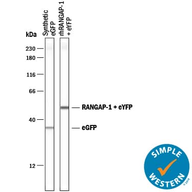
Detection of GFP by Simple WesternTM. Simple Western lane view shows eGFP and recombinant human RANGAP-1 tagged with eYFP, loaded at 0.2 mg/mL. Specific bands were detected for GFP at approximately 35 kDa and recombinant human RANGAP-1 tagged with eYFP at approximately 51 kDa (as indicated) using 10 µg/mL of Mouse Anti-GFP Monoclonal Antibody (Catalog # MAB42402). This experiment was conducted under reducing conditions and using the 12-230 kDa separation system.
Preparation and Storage
- 12 months from date of receipt, -20 to -70 °C as supplied.
- 1 month, 2 to 8 °C under sterile conditions after reconstitution.
- 6 months, -20 to -70 °C under sterile conditions after reconstitution.
Background: GFP
Green fluorescent protein (GFP) is a 27 kDa protein originally isolated from the jellyfish victoria. In the presence of UV light (490-520 nm), it emits a green fluorescent color that can be used to pinpoint locations of various intracellular proteins. GFP is 238 amino acids (aa) in length. It is a globular monomer that has a tendency to dimerize. The monomer has the shape of a beta -barrel with a chromophore (aa 65-67) containing alpha -helix running up its center. +36 GFP is a superpositively charged GFP variant that can penetrate mammalian cells with potencies much greater than that of cationic peptides or modestly cationic engineered proteins. Low molar concentrations of the protein can be observed within minutes of exposure.
Product Datasheets
FAQs
No product specific FAQs exist for this product, however you may
View all Antibody FAQsIsotype Controls
Reconstitution Buffers
Secondary Antibodies
Reviews for GFP Antibody
There are currently no reviews for this product. Be the first to review GFP Antibody and earn rewards!
Have you used GFP Antibody?
Submit a review and receive an Amazon gift card.
$25/€18/£15/$25CAN/¥75 Yuan/¥2500 Yen for a review with an image
$10/€7/£6/$10 CAD/¥70 Yuan/¥1110 Yen for a review without an image


