Human MIA Antibody Summary
Gly25-Gln131
Accession # Q16674
Customers also Viewed
Applications
Please Note: Optimal dilutions should be determined by each laboratory for each application. General Protocols are available in the Technical Information section on our website.
Scientific Data
 View Larger
Detection of Human MIA by Western Blot. Western blot shows lysates of Hs 294T human melanoma cell line. PVDF membrane was probed with 1 µg/mL of Goat Anti-Human MIA Antigen Affinity-purified Polyclonal Antibody (Catalog # AF2050) followed by HRP-conjugated Anti-Goat IgG Secondary Antibody (Catalog # HAF019). A specific band was detected for MIA at approximately 11 kDa (as indicated). This experiment was conducted under reducing conditions and using Immunoblot Buffer Group 8.
 View Larger
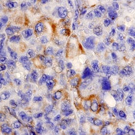
MIA in Human Melanoma. MIA was detected in immersion fixed paraffin-embedded sections of human melanoma using Goat Anti-Human MIA Antigen Affinity-purified Polyclonal Antibody (Catalog # AF2050) at 15 µg/mL overnight at 4 °C. Tissue was stained using the Anti-Goat HRP-DAB Cell & Tissue Staining Kit (brown; Catalog # CTS008) and counterstained with hematoxylin (blue). Specific staining was localized to plasma membranes of epithelial cells. View our protocol for Chromogenic IHC Staining of Paraffin-embedded Tissue Sections.
Preparation and Storage
- 12 months from date of receipt, -20 to -70 °C as supplied.
- 1 month, 2 to 8 °C under sterile conditions after reconstitution.
- 6 months, -20 to -70 °C under sterile conditions after reconstitution.
Background: MIA
MIA, also named Cartilage-Derived Retinoic Acid-Sensitive Protein (CD-RAP), is a secreted protein that plays an important role in melanoma metastasis. It is highly expressed in malignant melanomas and is associated with tumor progression.
Product Datasheets
Citations for Human MIA Antibody
R&D Systems personnel manually curate a database that contains references using R&D Systems products. The data collected includes not only links to publications in PubMed, but also provides information about sample types, species, and experimental conditions.
2
Citations: Showing 1 - 2
Filter your results:
Filter by:
-
Hypothalamic Rax+ tanycytes contribute to tissue repair and tumorigenesis upon oncogene activation in mice
Authors: W Mu, S Li, J Xu, X Guo, H Wu, Z Chen, L Qiao, G Helfer, F Lu, C Liu, QF Wu
Nature Communications, 2021-04-16;12(1):2288.
Species: Mouse
Sample Types: Whole Tissue
Applications: IHC -
Intracellular sortilin expression pattern regulates proNGF-induced naturally occurring cell death during development.
Authors: Nakamura K, Namekata K, Harada C, Harada T
Cell Death Differ., 2007-06-01;14(8):1552-4.
Species: Human
Sample Types: Tissue Homogenates, Whole Tissue
Applications: IHC, Western Blot
FAQs
No product specific FAQs exist for this product, however you may
View all Antibody FAQsIsotype Controls
Reconstitution Buffers
Secondary Antibodies
Reviews for Human MIA Antibody
Average Rating: 5 (Based on 1 Review)
Have you used Human MIA Antibody?
Submit a review and receive an Amazon gift card.
$25/€18/£15/$25CAN/¥75 Yuan/¥2500 Yen for a review with an image
$10/€7/£6/$10 CAD/¥70 Yuan/¥1110 Yen for a review without an image
Filter by:
AF2050 was tested along with MAB20501 in a sandwich assay. Although both antibodies worked as either the capture or detection, AF2050 had significantly higher affinity, which allowed quantification at a lower dilution of samples. The version of the assay we ended up using was AF2050 as both the capture and detection.


