Cytokines for Immune Cell Culture

Cytokines for Immune Cell Culture

Achieve Robust, Consistent Immune Cell Cultures with R&D Systems Cytokines

Immune cell culture is a key technique in immunology research for:

  • Investigating the functions of immune cells and their response to stimuli
  • Modeling immune responses to pathogens and diseases
  • Identifying cellular markers
  • Screening potential drugs
  • Expanding immune cells for the development of cell-based therapies

At R&D Systems, we recognize the importance of these studies, and we are committed to advancing immune cell research by developing and manufacturing proteins that will deliver superior performance and consistent results from one experiment to the next.

To support experimental needs from basic research to clinical applications, we offer research-grade proteins, Animal-free preclinical proteins, and GMP-grade proteins.

T Cells

CD4+ or CD8+ T cells that plays a central role in directing adaptive immune responses against invading pathogens.

CD4+ and CD8+ T cells play a central role in directing adaptive immune responses against invading microbial pathogens. Naïve CD4+ and CD8+ T cells are activated in a two-step process that requires: 1) recognition of an antigen/major histocompatibility complex (MHC) on an antigen-presenting cell (APC) by the T cell receptor (TCR), and 2) T cell co-stimulation mediated by co-stimulatory molecules on the APC interacting with specific T cell-expressed receptors. Following activation, naïve CD4+ and CD8+ T cells differentiate into one of several lineages of CD4+ T helper cell (Th) subsets or cytotoxic CD8+ T cell (Tc) subsets depending primarily on cytokines present in the extracellular environment. CD4+ and CD8+ T cells are subpopulations of CD3+ T cells, which account for 45-70% of peripheral blood mononuclear cells in healthy human adults. They are commonly isolated by immunomagnetic bead-based selection or by flow cytometry using cell surface markers. Cytokines such as IL-2, IL-7, and IL-15 are added to cultures to promote T cell proliferation and expansion, and the cytokines outlined below are used to promote in vitro differentiation of specific CD4+ T helper cell and CD8+ cytotoxic T cell subsets.


Cytokines for T Cell Culture or Differentiation

B7-H2/ICOS Ligand IFN-gamma* IL-1 beta* IL-2*† IL-4* IL-6*
IL-7*† IL-12 IL-15*† IL-18/IL-1F4 IL-21*† IL-23
IL-27 IL-33 TGF-beta 1* TNF-alpha* TSLP  
* GMP-grade proteins are available for these molecules.
† Liquid formulations of the GMP and Animal-free Preclinical grades of these proteins are available.

Detection of CD3+CD8+ and CD3+CD8- T cells by flow cytometry following enrichment in ExCellerate T Cell Expansion Media and R&D Systems cell culture cytokines.

Expansion of Human T Cells in ExCellerate™ Human T Cell Expansion Media Supplemented with Recombinant Human IL-2. Primary human peripheral blood mononuclear cells (PBMCs) were cultured for 9 days in ExCellerate Human T Cell Expansion Media (Catalog # CCM030) using activating beads and Recombinant Human IL-2 (Catalog # 202-IL). (A) Cell counts were performed to determine the fold expansion compared to the Day 0 seeding density of 0.25 x 10⁶ cells/mL. (B) T cells enriched during the culture were detected by staining with an APC-conjugated Mouse Anti-Human CD3 epsilon Monoclonal Antibody (Catalog # FAB100A) and with a PE-conjugated Mouse Anti-Human CD8 alpha Monoclonal Antibody (Catalog # FAB1509P).

Cytokines for Differentiating CD4+ T Helper Cell Subsets

Cytokines for Differentiating CD8+ Cytotoxic T Cell Subsets

Tc1 Tc2 Tc9 Tc17 Tc22 Tfc Treg
IL-2 IL-4 IL-4 IL-6 IL-6 IL-6 TGF-beta 1
IL-12   TGF-beta 1 IL-21 TNF-alpha IL-21  
      TGF-beta 1   IL-23  
          TGF-beta 1  

Natural Killer Cells

Natural killer cell with both cytotoxic and cytokine-secreting capabilities can be expanded and activated with R&D Systems cell culture cytokines.

Natural killer (NK) cells are innate lymphoid cells that play a key role in the early response to infection or malignant transformation. NK cells have both cytotoxic activity and cytokine-secreting capabilities. Their cytotoxic activity enables NK cells to directly destroy target cells through the release of cytolytic granules, while their cytokine-secreting capabilities allow them to regulate downstream immune responses. NK cell activation is controlled by the integration of signals received from multiple cell surface receptors that deliver either activating or inhibitory signals. Under normal physiological conditions, NK cell activation is inhibited by ligands expressed on healthy cells that engage inhibitory receptors on NK cells. Detection of abnormalities in infected or cancerous cells, such as the down-regulation of MHC class I expression or the elevated expression of stress-induced ligands, leads to NK cell activation. NK cells make up
5-20% of peripheral blood mononuclear cells and a variety of cytokines, including IL-2, IL-12, IL-15, IL-18, and IL-21, have been used for their in vitro expansion, differentiation, and activation.

Cytokines for NK Cell Culture and Differentiation

IL-2*† IL-12 IL-15*† IL-18/IL-1F4 IL-21*†

* GMP-grade proteins are available for these molecules.

† Liquid formulations of the GMP and Animal-free Preclinical grades of these proteins are available.

Characterization of human NK cells on days 0 and 13 of culture with R&D Systems cytokines indicates a CD3+CD56+GranzymeK+GranzymeB+Perforin+ phenotype on day 13 by flow cytometry.

Culture and Characterization of Human Natural Killer Cells. Natural killer cells were expanded from peripheral blood mononuclear cells (PBMCs) using plate-bound Anti-Human NKp46 Monoclonal Antibody (Catalog # MAB1850) in ExCellerate Human NK Cell Expansion Media, Xeno-Free (Catalog # CCM032) plus Recombinant Human IL-2 (Catalog # 202-IL; 27 ng/mL), Recombinant Human IL-12 (Catalog # 219-IL; 10 ng/mL), Recombinant Human IL-18/IL-1F4 (Catalog # 9124-IL; 10 ng/mL), and Recombinant Human IL-21 (Catalog # 8879-IL; 10 ng/mL) for 13 days. NK cells were assessed on Days 0 and 13 using an Alexa Fluor® 647-conjugated Mouse Anti-Human CD56 Monoclonal Antibody (Catalog # FAB24086R), an Alexa Fluor® 405-conjugated Mouse Anti-Human CD3 Monoclonal Antibody (Catalog # FAB100V), a PerCP-conjugated Mouse Anti-Human CD16 Monoclonal Antibody (Catalog # FAB2546C), an Alexa Fluor® 594-conjugated Rabbit Anti-Human Granzyme K Monoclonal Antibody, an Alexa Fluor® 750-conjugated Mouse Anti-Human Perforin Monoclonal Antibody, and an Alexa Fluor® 488-conjugated Mouse Anti-Human Granzyme B Monoclonal Antibody (Catalog # IC2906G). Cells were gated on singlets (FSC-A by FSC-H) and live cells and quadrants were set using isotype controls (not shown). Alexa Fluor® is a registered trademark of Molecular Probes, Inc., Eugene, OR.

Monocytes/Macrophages

Monocytes can be differentiated into macrophages or dendritic cells using R&D Systems cell culture cytokines.

Monocytes are myeloid cells that along with dendritic cells and macrophages, comprise the mononuclear phagocyte system. Cells belonging to this system have many common features including the ability to engulf and digest large particles, such as bacteria, fungi, viruses, and dying cells, and the ability to function as antigen-presenting cells. Additionally, monocytes, macrophages, and dendritic cells can secrete cytokines and chemokines that direct the migration and functions of other immune cell types. Monocytes circulate in the blood and migrate to sites of tissue inflammation, where they can differentiate into macrophages or dendritic cells. Monocytes typically account for 10-20% of peripheral blood mononuclear cells in humans and can be isolated by plastic adhesion, magnetic bead-based negative selection, or CD14+ positive selection. Following their isolation, CD14+ monocytes can be differentiated into pro-inflammatory M1 or anti-inflammatory M2 macrophages in the presence of GM-CSF or M-CSF, respectively. Microbial products and pro-inflammatory cytokines such as lipopolysaccharide, IFN-γ, and TNF-α are used to drive classical M1 macrophage activation, while IL-4 and IL-13 are commonly used to promote alternative M2 macrophage activation.

Cytokines for Monocyte/Macrophage Culture and Differentiation

GM-CSF* IFN-gamma* IL-1 beta* IL-4* IL-10* IL-13
M-CSF* TNF-alpha*      
* GMP-grade proteins are available for these molecules.

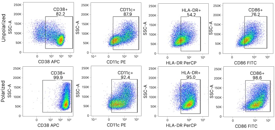
M1-activated macrophages cultured using R&D Systems cytokines and LPS display a CD38+CD11c+HLA-DR+CD86+ phenotype as determined by flow cytometry after 4 days.

Culture and Characterization of M1-Activated Human Macrophages. Monocytes were isolated from peripheral blood mononuclear cells (PBMCs) via adherence depletion for 5 hours. Non-adherent cells were removed. Adherent cells were incubated for 6 days in C3 + 10% huAB media with Recombinant Human GM-CSF (Catalog # 215-GM; 50 ng/mL). Media and cytokines were refreshed on day 3. For M1 polarization, cells were incubated with Recombinant Human GM-CSF (Catalog # 215-GM; 50 ng/mL), Recombinant Human IFN-γ (Catalog # 285-IF; 50 ng/mL), and LPS (50 ng/mL) 24 hours prior to staining. Cells were stained with an Alexa Fluor® 405-conjugated Mouse Anti-Human CD3 Monoclonal Antibody (Catalog # FAB100V), an Alexa Fluor® 405-conjugated Mouse Anti-Human CD20 Monoclonal Antibody (Catalog # FAB4225V), and an Alexa Fluor® 700-conjugated Mouse Anti-Human CD56 Monoclonal Antibody (Catalog # FAB24086N), and live CD3-CD20-CD56- cells were detected and gated by flow cytometry. M1-activated macrophages were identified by staining with an APC-conjugated Mouse Anti-Human CD38 Monoclonal Antibody (Catalog # FAB2404A), a PE-conjugated Mouse Anti-Human CD11c Monoclonal Antibody (Catalog # FAB1777P), a PerCP-conjugated Mouse Anti-Human HLA-DR Monoclonal Antibody (Catalog # FAB4869C), and a FITC-conjugated Mouse Anti-Human CD86/B7-2 Monoclonal Antibody (Catalog # FAB141F). Alexa Fluor® is a registered trademark of Molecular Probes, Inc., Eugene, OR.

Dendritic Cells

M1-activated macrophages cultured using R&D Systems cytokines and LPS display a CD38+CD11c+HLA-DR+CD86+ phenotype as determined by flow cytometry after 4 days.

Dendritic cells are professional antigen-presenting cells that promote CD4+ and CD8+ T cell activation. They play a central role in innate and adaptive immune responses due to their abilities to recognize, phagocytose, and process antigens from invading pathogens, upregulate MHC molecules and co-stimulatory molecules, and travel to the secondary lymphoid organs to prime T cell activation. In addition, dendritic cells produce polarizing cytokines that direct the differentiation of pathogen-specific effector T cell subsets. Dendritic cells are present in the skin, mucosal surfaces, peripheral blood, and lymphoid and non-lymphoid tissues, where they continuously sample the extracellular environment for foreign particles and differentially shape the immune response depending on the type of pathogen detected. Significantly, dendritic cells also play a key role in promoting immune homeostasis by continually presenting T cells with self-antigens to promote self-tolerance and secreting tolerogenic cytokines that induce regulatory T cell differentiation. Dendritic cells can be isolated from human blood mononuclear cells or differentiated from CD14+ monocytes or embryonic stem cells. Common cytokines used for differentiating monocyte-derived dendritic cells include GM-CSF and IL-4. Additional cytokines, such as TNF-α are used to drive dendritic cell maturation.

Cytokines for Dendritic Cell Culture, Activation, and Maturation

Flt-3 Ligand/FLT3L* GM-CSF* IFN-alpha IFN-gamma* IL-1beta* IL-3*
IL-4* IL-6* IL-12 M-CSF* SCF* TNF-alpha*
* GMP-grade proteins are available for these molecules.

Birghtfield image of immature dendritic cells following a 7 day differentiation of CD14+ monocytes with R&D Systems cell culture cytokines.

Morphology of Immature Dendritic Cells Differentiated from CD14+ Monocytes. Immature dendritic cells were generated from human CD14+ enriched peripheral blood mononuclear cells (PBMCs) following culture for seven days in StemXVivo™ Serum-Free Dendritic Cell Base Media (Catalog # CCM003) supplemented with Recombinant Human IL-4 (Catalog # 204-IL) and Recombinant Human GM-CSF (Catalog # 215-GM).

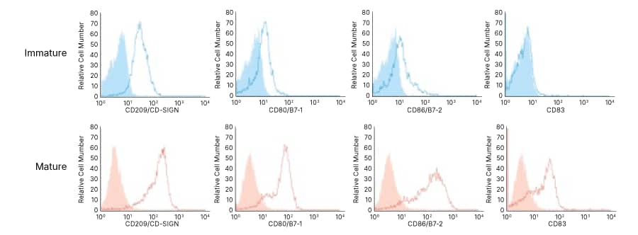
Flow cytometry analysis comparing immature and LPS-treated mature MoDCs differentiated from CD14+ monocytes using R&D Systems cell culture cytokines.

Phenotypic Analysis of Cultured Immature and Mature Monocyte-derived Dendritic Cells. Immature monocyte-derived dendritic cells (top) were obtained after CD14+-enriched monocytes were cultured for seven days in StemXVivo Serum-Free Dendritic Cell Base Media (Catalog # CCM003) supplemented with Recombinant Human IL-4 (Catalog # 204-IL) and Recombinant Human GM-CSF (Catalog # 215-GM). Mature monocyte-derived dendritic cells (bottom) were obtained by culturing CD14+-enriched monocytes under the same conditions for seven days and then treating the cells with Recombinant Human TNF-α (Catalog # 210-TA) for an additional 3 days. The phenotypes of Day 7 immature monocyte-derived dendritic cells and day ten TNF-α-treated mature monocyte-derived dendritic cells were assessed by staining with a Mouse Anti-Human DC-SIGN/CD209 Monoclonal Antibody (Catalog # MAB161), a Mouse Anti-Human CD80/B7-1 Monoclonal Antibody (Catalog # MAB140), a Mouse Anti-Human CD86/B7-2 Monoclonal Antibody (Catalog # MAB141), or a Mouse Anti-Human CD83 Monoclonal Antibody (Catalog # MAB1774) and a PE-conjugated Goat Anti-Mouse IgG Polyclonal Antibody (Catalog # F0102B; open histograms), or an appropriate isotype control antibody (filled histograms). Stained cells were analyzed by flow cytometry.

B Cells

B cells can be expanded or differentiated using R&D Systems cell culture cytokines such as IL-4, CD40 Ligand, or anti-CD40.

B cells are essential components of the adaptive immune response due to their ability to produce antibodies against foreign antigens and generate immunological memory. Additionally, B cells can function as antigen-presenting cells and secrete immunomodulatory cytokines that regulate the activities of other immune cell types and lymphoid tissue organization. B cells represent 5-10% of peripheral blood mononuclear cells in humans and are commonly enriched for using magnetic bead-based negative selection, or CD19+ positive selection. B cells can be further expanded in vitro in the presence of IL-4, CD40 Ligand, or anti-CD40. Additional cytokines can be used to promote B cell differentiation, antibody production, or enhance the production of specific antibody isotypes such as IgA, IgG, IgE, or IgM.

Cytokines for B Cell Culture and Differentiation

BAFF/BLyS/TNFSF13B CD40 Ligand IL-2*† IL-4* IL-5 IL-10*
IL-15*† IL-21* RANK Ligand TNF-alpha*  
* GMP-grade proteins are available for these molecules.
† Liquid formulations of the GMP and Animal-free Preclinical grades of these proteins are available.

A:Graph showing robust expansion of mouse B cells in ExCellerate media with IL-4 and Anti-Mouse CD40 Antibody
B: Brightfield image of mouse B cell colonies expanded in ExCellerate media with IL-4 and Anti-Mouse CD40 Antibody 
C:Flow cytometry analysis of mouse B cells expanded in ExCellerate media show >98% are B220<sup>+</sup>CD19<sup>+</sup> and CD3-CD4-

Expansion of Mouse B Cells in ExCellerate Human B Cell Expansion Media Supplemented with Recombinant Mouse IL-4 and an Anti-Mouse CD40 Antibody. Mouse B cells were isolated from splenocytes using the MagCellect™ Mouse B Cell Isolation Kit (Catalog # MAGM204) and expanded using ExCellerate B Cell Media (Catalog # CCM031) with or without Recombinant Mouse IL-4 (Catalog # 404-ML) and a Rat Anti-Mouse CD40/TNFRSF5 Monoclonal Antibody (Catalog # MAB440). (A) Expansion of mouse B cells was monitored using Rezasurin (Catalog # AR002). Data show that ExCellerate B Cell Media supplemented with IL-4 and an Anti-Mouse CD40 Antibody results in robust mouse B cell expansion. (B) Representative brightfield images of mouse B cell colonies. (C) Expanded mouse B cells are B220+CD19+ (>98%) and negative for both CD3 and CD4.

Bulk Protein banner

Benefits of R&D Systems Proteins for Immune Cell Culture

  • High Bioactivity and Performance: Utilizing over 900 different bioassays, the biological activity of every protein we offer is tested in an appropriate bioassay to confirm that it meets our strict QC activity parameters.
  • Lot-to-Lot Consistency: Minimal lot-to-lot variability is ensured by testing each new lot side-by-side with previous lots, so you don’t have to worry whether results will be reproducible over time.
  • High Purity and Low Endotoxin Levels: Our proteins are typically over 95% pure and have a guaranteed industry-leading endotoxin level of 0.1 EU/ug by the LAL method.
  • Seamless Transition from Preclinical Research to Clinical Manufacturing: Our Animal-free RUO and GMP-grade proteins originate from the same clone, sequence, and expression system to make the transition from preclinical research into clinical manufacturing as seamless as possible.
  • Supply Chain Reliability: We have the capacity and the experience to ensure that we can provide a stable supply of the proteins needed for your research.
  • Bulk Proteins at Discounted Prices: Unlock competitive discounts on bulk protein orders of 1 mg or more with our tiered pricing model. The more protein you need, the more you save.

Antibodies for Immune Cell Activation and Differentiation

In addition to our recombinant proteins for immune cell culture, we offer a wide selection of research use only or GMP antibodies to optimize your immune cell activation and differentiation protocols. Our antibodies undergo extensive in-house validation to ensure performance, and we provide data images, citations, reviews, and a 100% guarantee for each of our antibody products, so you can have confidence they will work in the applications and species listed on our datasheets.

CD3* CD28* CD40 Fc gamma RIII/CD16 IFN-gamma IL-4
IL-12 NKp30 NKp46      
* GMP antibodies are available for these molecules.

Benefits of R&D Systems Antibodies

  • Guaranteed Performance: Data images, citations, reviews, and a 100% satisfaction guarantee enable customers to have confidence that our antibodies will work in the applications and species designated.
  • Extensive Validation and Product Support: Extensive in-house validation and expert product support are provided to ensure the performance of our antibodies.

Additional Products to Improve Immune Cell Culture Consistency

ExCellerate™ Media for Immune Cell Culture

Provides an optimized serum-free culture environment for the generation and expansion of individual immune cell types.

Animal-free Recombinant Proteins

Provides a seamless transition from preclinical research into clinical manufacturing, ensures defined culture conditions, and eliminates regulatory or ethical concerns associated with animal-derived reagents. Our Animal-free proteins are produced using identical expression systems and manufacturing methods as our Animal-free GMP-grade proteins.

GMP Proteins

Manufactured under guidelines that allow for their use as ancillary materials in cell therapy manufacturing processes. These proteins undergo extensive quality control testing and come with comprehensive documentation and full transparency and traceability of source and manufacturing system. Liquid, process-sized, and closed process options are also available for GMP cytokines. Learn more.

Cell Expansion and Differentiation Kits

Optimized reagents for the differentiation and expansion of leukocyte populations in culture.

Isolation Kits

Positive and negative cell selection kits to isolate immune cells for cell culture.

Cell Proliferation and Viability Assays

Reagents for studying cell proliferation, viability, and cytotoxicity including fluorescent reporter dyes and MTT assays.