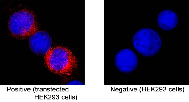
Mouse Ret Antibody Summary
Leu29-Arg637 (Phe174Ser)
Accession # P35546
Customers also Viewed
Applications
Please Note: Optimal dilutions should be determined by each laboratory for each application. General Protocols are available in the Technical Information section on our website.
Scientific Data
 View Larger
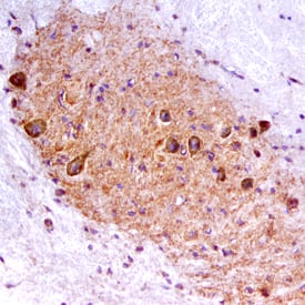
Ret in Mouse Spinal Cord. Ret was detected in perfusion fixed frozen sections of mouse spinal cord using Goat Anti-Mouse Ret Antigen Affinity-purified Polyclonal Antibody (Catalog # AF482) at 15 µg/mL overnight at 4 °C. Tissue was stained using the Anti-Goat HRP-DAB Cell & Tissue Staining Kit (brown; Catalog # CTS008) and counterstained with hematoxylin (blue). Specific staining was localized to the ventral horn. View our protocol for Chromogenic IHC Staining of Frozen Tissue Sections.
 View Larger
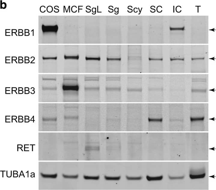
Detection of Porcine Ret by Western Blot ERBB-family signaling molecules in rat testis cells. (a) Polypeptides in the EGF super-family signal by activating ERBB-family transmembrane receptor tyrosine kinases. ERBB1 is a receptor for ‘classical’ low molecular weight EGF-like peptides. ERBB2 is the primary transducer for ligand-bound ERBB1, ERBB3 and ERBB4. ERBB2’s extracellular domain does not bind known ligands. ERBB3 is a receptor for Neuregulin-1 (NRG1), NRG2 and Neuroglycan-C (CSPG5). Ligand bound ERBB3 displays poor kinase activity and signals most effectively as a heteromer with ERBB1, ERBB2 and/or ERBB4. ERBB4 is a receptor for NRG1, NRG2, NRG3 and NRG4 plus other EGF-like peptides*. (b) Western blotting analysis of ERBB-family proteins in fractions of testis cells from 23-day-old rats. Lysates of type A spermatogonia after proliferating for ~180 days/15 passages in culture (SgL), freshly isolated laminin-binding type A spermatogonia (Sg), laminin non-binding spermatogenic cells (Scy), tubular somatic cells (SC), interstitial somatic cells (IC), MCF7 human mammary gland cells (MCF) and COS7 monkey kidney cells (COS). Arrowheads: ERBBs 1–4 (~185 kDa), RET (~155 and 170 kDa) and TUBA1a (~55 kDa). (c) Relative abundance (qtPCR) of ERBB-family transcripts in testis cells isolated from 23-day-old rats (n=cells from three different rats; ±S.E.M.). Spermatogonia (Sg), Spermatocytes (Scy; differentiating spermatogonia/early spermatocytes), Tubular somatic cells (SC) and Interstitial somatic cells (IC) are cell types described in panel (b). (d) Testis cross-section from 26-day-old tgGCS-EGFP transgenic rats labeled with anti-ERBB2 (Red) overlaying EGFP fluorescence from germ cells (green). Note, cytoplasmic ERBB2 labeling in germ cells resembling differentiating spermatogonia (white arrows) and spermatocytes (yellow arrow). Scale, 40 μm. (e) Rat seminiferous tubule whole mount from 24-day-old wild-type rat labeled using antibodies to ERBB2 (Red) and ZBTB16 (Green). Scale, 20 μm. Note: nuclear ZBTB16 labeling is more robust in ERBB2-dim spermatogonia (cyan arrows), compared with ERBB2-bright spermatogenic cells (white arrows). (f) Rat seminiferous tubule whole mount from a 24-day-old wild-type rat labeled with antibodies to ERBB2 (Red) and phospho-Histone-3 (pH3, Green). Scale, 40 μm. Note: nuclear pH3 in large mitotic ERBB2+ syncytia. Image collected and cropped by CiteAb from the following open publication (https://www.nature.com/articles/cddiscovery201518), licensed under a CC-BY license. Not internally tested by R&D Systems.
 View Larger
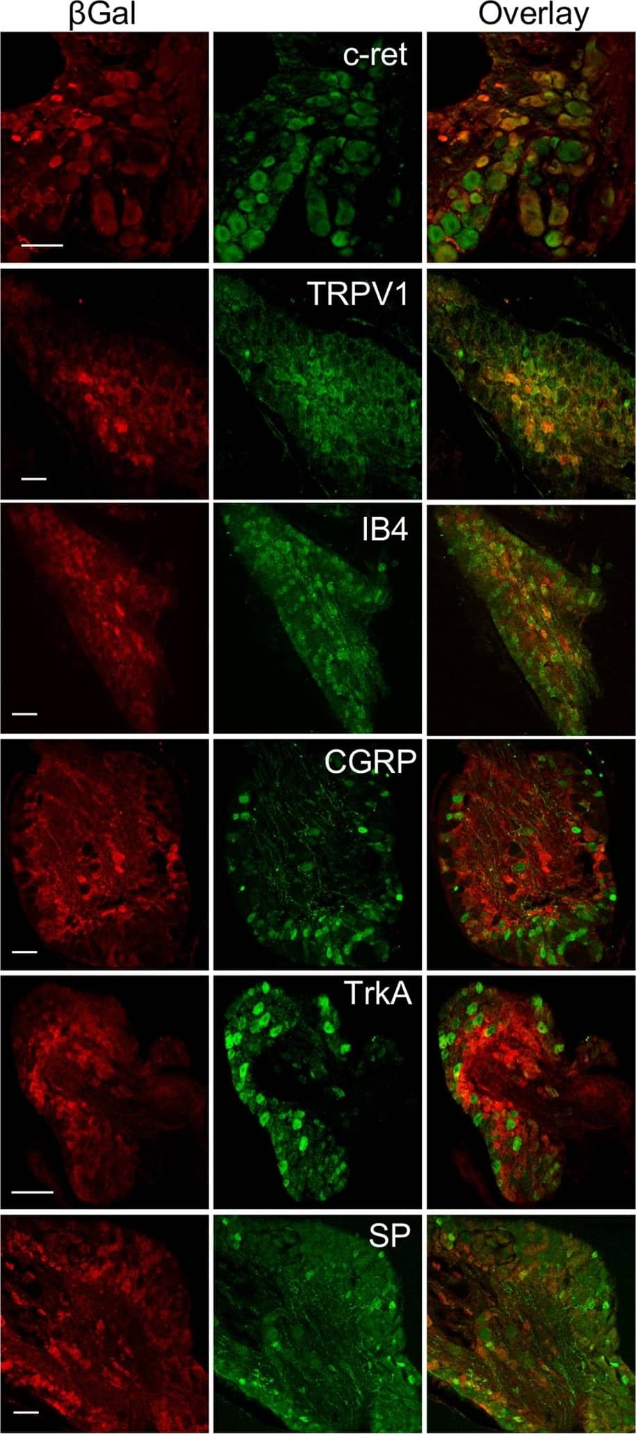
Detection of Mouse Ret by Immunohistochemistry P2RX4 expression in Dorsal Root Ganglion neurons. (A) Representative cropped western blotting analysis of lumbar DRG extract indicates the presence of a 60 kDa band in P2RX4+/+ DRG that is absent in extracts from P2RX4−/− DRG. N = 3 independent experiments, n = 3 mice per genotype. (B) beta -galactosidase staining was used as a surrogate marker for P2RX4 in DRG sections. Only P2RX4−/− neurons are immuno-positive for beta gal compared to sections from P2RX4+/+ DRG. Scale bar 100 µm. (C) DRG neurons that express beta -gal innervate paw skin. Fluoro-Gold B, a retrograde marker of neurons was injected in paw-skin and L5/L6 DRG were removed for staining a week later. beta gal immunostaining reveals a population of neurons that are labeled for both beta gal and Fluoro-Gold B. Scale bar 50 µm. (D) In nociceptive neurons, beta gal is mainly expressed in c-ret-, TRPV1- and IB4-positive neurons, but scarcely in others populations (CGRP, TrkA or Substance P (SP) expressing cells). Representative images of DRG sections stained with beta gal and makers of nociceptive neurons. Scale bars 50 µm. Image collected and cropped by CiteAb from the following open publication (https://pubmed.ncbi.nlm.nih.gov/29343707), licensed under a CC-BY license. Not internally tested by R&D Systems.
 View Larger
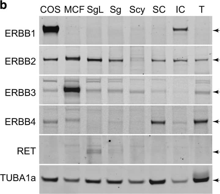
Detection of Rat Ret by Western Blot ERBB-family signaling molecules in rat testis cells. (a) Polypeptides in the EGF super-family signal by activating ERBB-family transmembrane receptor tyrosine kinases. ERBB1 is a receptor for ‘classical’ low molecular weight EGF-like peptides. ERBB2 is the primary transducer for ligand-bound ERBB1, ERBB3 and ERBB4. ERBB2’s extracellular domain does not bind known ligands. ERBB3 is a receptor for Neuregulin-1 (NRG1), NRG2 and Neuroglycan-C (CSPG5). Ligand bound ERBB3 displays poor kinase activity and signals most effectively as a heteromer with ERBB1, ERBB2 and/or ERBB4. ERBB4 is a receptor for NRG1, NRG2, NRG3 and NRG4 plus other EGF-like peptides*. (b) Western blotting analysis of ERBB-family proteins in fractions of testis cells from 23-day-old rats. Lysates of type A spermatogonia after proliferating for ~180 days/15 passages in culture (SgL), freshly isolated laminin-binding type A spermatogonia (Sg), laminin non-binding spermatogenic cells (Scy), tubular somatic cells (SC), interstitial somatic cells (IC), MCF7 human mammary gland cells (MCF) and COS7 monkey kidney cells (COS). Arrowheads: ERBBs 1–4 (~185 kDa), RET (~155 and 170 kDa) and TUBA1a (~55 kDa). (c) Relative abundance (qtPCR) of ERBB-family transcripts in testis cells isolated from 23-day-old rats (n=cells from three different rats; ±S.E.M.). Spermatogonia (Sg), Spermatocytes (Scy; differentiating spermatogonia/early spermatocytes), Tubular somatic cells (SC) and Interstitial somatic cells (IC) are cell types described in panel (b). (d) Testis cross-section from 26-day-old tgGCS-EGFP transgenic rats labeled with anti-ERBB2 (Red) overlaying EGFP fluorescence from germ cells (green). Note, cytoplasmic ERBB2 labeling in germ cells resembling differentiating spermatogonia (white arrows) and spermatocytes (yellow arrow). Scale, 40 μm. (e) Rat seminiferous tubule whole mount from 24-day-old wild-type rat labeled using antibodies to ERBB2 (Red) and ZBTB16 (Green). Scale, 20 μm. Note: nuclear ZBTB16 labeling is more robust in ERBB2-dim spermatogonia (cyan arrows), compared with ERBB2-bright spermatogenic cells (white arrows). (f) Rat seminiferous tubule whole mount from a 24-day-old wild-type rat labeled with antibodies to ERBB2 (Red) and phospho-Histone-3 (pH3, Green). Scale, 40 μm. Note: nuclear pH3 in large mitotic ERBB2+ syncytia. Image collected and cropped by CiteAb from the following open publication (https://www.nature.com/articles/cddiscovery201518), licensed under a CC-BY license. Not internally tested by R&D Systems.
 View Larger
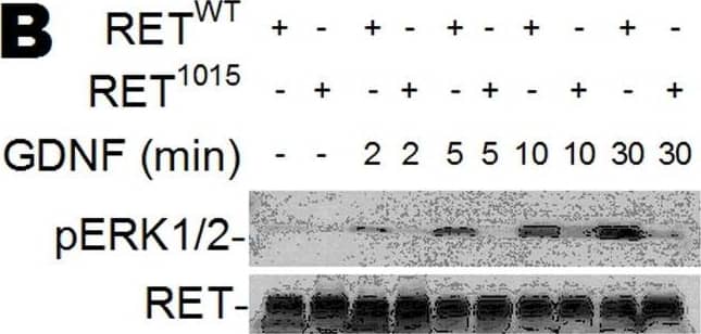
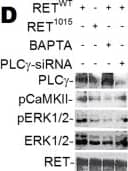
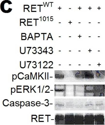
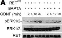
Detection of Mouse Ret by Western Blot GDNF-induced Ca2+ signaling phosphorylates ERK1/2 and CaMKII.(A–D) Western blot of HeLa cells transfected with RETWT or RET1015 treated with GDNF (100 ng/ml). GDNF triggers time dependent phosphorylation of ERK1/2 (pERK1/2) in RETWT cells that is suppressed by BAPTA (10 µM) (A). Less pERK1/2 is observed in cells transfected with RET1015 than RETWT (B). GDNF-induced phosphorylation of CaMKII (pCaMKII) or pERK1/2 is suppressed when blocking PLC with U73122 (5 µM) (C) or knocking down PLC gamma with siRNA (PLC gamma -siRNA) (D). Treating RETWT cells with the U73122 analogue U73343 (5 µM) had no effect on GDNF-activated pCaMKII or pERK1/2 (C). Increased Caspase-3 cleavage was not detected in cells treated with the inhibitors BAPTA or U73122 (C). Image collected and cropped by CiteAb from the following open publication (https://pubmed.ncbi.nlm.nih.gov/22355350), licensed under a CC-BY license. Not internally tested by R&D Systems.
Preparation and Storage
- 12 months from date of receipt, -20 to -70 °C as supplied.
- 1 month, 2 to 8 °C under sterile conditions after reconstitution.
- 6 months, -20 to -70 °C under sterile conditions after reconstitution.
Background: Ret
The GDNF family of neurotrophic factors consititute a new family of factors within the TGF-beta superfamily. These proteins are potent survival factors for various central and peripheral neurons during development and in the adult animal. The GDNF family members (GDNF, neurturin and persephin) signal through multicomponent receptors that consist of the Ret receptor tyrosine kinase and one of four glycosyl-phosphatidylinositol (GPI)-linked ligand-binding subunits (GFR alpha ‑1 ‑ 4). GFR alpha -1, -2, and -4 are the preferred ligand-binding subunits for GDNF, neurturin and persephin, respectively. To date, the preferred ligand for GFR alpha ‑3 has not been identified. The Ret tyrosine-kinase receptor is encoded by the c-ret proto-oncogene. Mutations of the ret gene have been associated with various human diseases affecting tissues derived from the neural crest, including Hirschsprung’s disease, multiple endocrine neoplasia MEN2A and MEN2B, and familial medullary thyroid carcinoma. Mouse Ret cDNA encodes a 1115 amino acid (aa) residue transmembrane tyrosine kinase with a 28 aa residue signal peptide, a 609 aa residue cysteine-rich extracellular domain and a 456 aa residue cytoplasmic domain. A cadherin-related sequence is also present in the extracellular domain of Ret. Human and mouse Ret share 83% amino acid sequence homology (77% homology in the extracellular domain and 93% homology in the cytoplasmic domain). Although Ret does not bind GDNF ligands directly, the extracellular domain of Ret binds the GDNF-GFR-alpha complex with high affinity and is a potent GDNF antagonist in the presence of soluble GFR-alpha.
- Trupp, M. et al. (1998) Mol. Cell Neurosci. 11:47.
- Enokido, Y. et al. (1998) Curr. Biol. 8:1019.
- Carlomagno, F. et al. (1998) Endocrinology, 139:3613.
Product Datasheets
Citations for Mouse Ret Antibody
R&D Systems personnel manually curate a database that contains references using R&D Systems products. The data collected includes not only links to publications in PubMed, but also provides information about sample types, species, and experimental conditions.
40
Citations: Showing 1 - 10
Filter your results:
Filter by:
-
Boundary cap neural crest stem cells homotopically implanted to the injured dorsal root transitional zone give rise to different types of neurons and glia in adult rodents.
Authors: Trolle, Carl, Konig, Niclas, Abrahamsson, Ninnie, Vasylovska, Svitlana, Kozlova, Elena N
BMC Neurosci, 2014-05-05;15(0):60.
-
Expression of Glial Cell‐Derived Neurotrophic Factor Receptors Within Nucleus Ambiguus During Rat Development
Authors: Quinton Blount, Ignacio Hernandez‐Morato, Yalda Moayedi, Michael J. Pitman
The Laryngoscope
-
BMSCs Promote Differentiation of Enteric Neural Precursor Cells to Maintain Neuronal Homeostasis in Mice With Enteric Nerve Injury
Authors: Mengke Fan, Huiying Shi, Hailing Yao, Weijun Wang, Yurui Zhang, Chen Jiang et al.
Cellular and Molecular Gastroenterology and Hepatology
-
The impact of C-tactile low-threshold mechanoreceptors on affective touch and social interactions in mice
Authors: D Huzard, M Martin, F Maingret, J Chemin, F Jeanneteau, PF Mery, P Fossat, E Bourinet, A François
Oncogene, 2022-06-29;8(26):eabo7566.
Species: Mouse
Sample Types: Whole Tissue
Applications: IHC -
Single cell RNA sequencing identifies early diversity of sensory neurons forming via bi-potential intermediates
Authors: L Faure, Y Wang, ME Kastriti, P Fontanet, KKY Cheung, C Petitpré, H Wu, LL Sun, K Runge, L Croci, MA Landy, HC Lai, GG Consalez, A de Chevign, F Lallemend, I Adameyko, S Hadjab
Nat Commun, 2020-08-21;11(1):4175.
Species: Mouse
Sample Types: Whole Tissue
Applications: IHC -
NOP receptor agonist attenuates nitroglycerin-induced migraine-like symptoms in mice
Authors: Katarzyna M. Targowska-Duda, Akihiko Ozawa, Zachariah Bertels, Andrea Cippitelli, Jason L. Marcus, Hanna K. Mielke-Maday et al.
Neuropharmacology
-
Identification of Spinal Neurons Contributing to the Dorsal Column Projection Mediating Fine Touch and Corrective Motor Movements
Authors: Sónia Paixão, Laura Loschek, Louise Gaitanos, Pilar Alcalà Alcalà Morales, Martyn Goulding, Rüdiger Klein
Neuron
-
Muscle-selective RUNX3 dependence of sensorimotor circuit development
Authors: Yiqiao Wang, Haohao Wu, Pavel Zelenin, Paula Fontanet, Simone Wanderoy, Charles Petitpré et al.
Development
-
A cell fitness selection model for neuronal survival during development
Authors: Y Wang, H Wu, P Fontanet, S Codeluppi, N Akkuratova, C Petitpré, Y Xue-Franzé, K Niederreit, A Sharma, F Da Silva, G Comai, G Agirman, D Palumberi, S Linnarsson, I Adameyko, A Moqrich, A Schedl, G La Manno, S Hadjab, F Lallemend
Nat Commun, 2019-09-12;10(1):4137.
Species: Mouse
Sample Types: Whole Cells
Applications: ICC -
Spinal Neuropeptide Y1 Receptor-Expressing Neurons Form an Essential Excitatory Pathway for Mechanical Itch
Authors: D Acton, X Ren, S Di Costanz, A Dalet, S Bourane, I Bertocchi, C Eva, M Goulding
Cell Rep, 2019-07-16;28(3):625-639.e6.
Species: Mouse
Sample Types: Whole Tissue
Applications: IHC-Fr -
PRDM12 Is Required for Initiation of the Nociceptive Neuron Lineage during Neurogenesis
Authors: L Bartesaghi, Y Wang, P Fontanet, S Wanderoy, F Berger, H Wu, N Akkuratova, F Bouçanova, JJ Médard, C Petitpré, MA Landy, MD Zhang, P Harrer, C Stendel, R Stucka, M Dusl, ME Kastriti, L Croci, HC Lai, GG Consalez, A Pattyn, P Ernfors, J Senderek, I Adameyko, F Lallemend, S Hadjab, R Chrast
Cell Rep, 2019-03-26;26(13):3484-3492.e4.
Species: Mouse
Sample Types: Whole Tissue
Applications: IHC -
Non-canonical Ret signaling augments p75-mediated cell death in developing sympathetic neurons
Authors: Christopher R. Donnelly, Nicole A. Gabreski, Esther B. Suh, Monzurul Chowdhury, Brian A. Pierchala
Journal of Cell Biology
-
Tumor necrosis factor-? mediated pain hypersensitivity through Ret receptor in resiniferatoxin neuropathy
Authors: SC Lu, YS Chang, HW Kan, YL Hsieh
Kaohsiung J. Med. Sci., 2018-09-01;34(9):494-502.
Species: Mouse
Sample Types: Whole Tissue
Applications: IHC-P -
RET-mediated glial cell line derived neurotrophic factor signaling inhibits mouse prostate development
Authors: HJ Park, EC Bolton
Development, 2017-05-15;0(0):.
Species: Mouse
Sample Types: Tissue Homogenates, Whole Tissue
Applications: IHC, Western Blot -
Neurturin and a Glp-1 Analogue Act Synergistically to Alleviate Diabetes in Zucker Diabetic Fatty Rats
Authors: JL Trevaskis, CB Sacramento, H Jouihan, S Ali, J Le Lay, S Oldham, N Bhagroo, BB Boland, J Cann, Y Chang, T O'Day, V Howard, C Reers, MS Winzell, DM Smith, M Feigh, P Barkholt, K Schreiter, M Austen, U Andag, S Thompson, L Jermutus, MP Coghlan, J Grimsby, C Dohrmann, CJ Rhodes, CM Rondinone, A Sharma
Diabetes, 2017-04-13;0(0):.
Species: Rat
Sample Types: Whole Tissue
Applications: IHC -
Genetic ablation of GINIP-expressing primary sensory neurons strongly impairs Formalin-evoked pain
Authors: L Urien, S Gaillard, L Lo Re, P Malapert, M Bohic, A Reynders, A Moqrich
Sci Rep, 2017-02-27;7(0):43493.
Species: Mouse
Sample Types: Whole Tissue
Applications: IHC-Fr -
A Brainstem-Spinal Cord Inhibitory Circuit for Mechanical Pain Modulation by GABA and Enkephalins
Authors: Amaury François, Sarah A. Low, Elizabeth I. Sypek, Amelia J. Christensen, Chaudy Sotoudeh, Kevin T. Beier et al.
Neuron
-
Exon Skipping in the RET Gene Encodes Novel Isoforms That Differentially Regulate RET Protein Signal Transduction
Authors: NA Gabreski, JK Vaghasia, SS Novakova, NQ McDonald, BA Pierchala
J. Biol. Chem., 2016-05-23;291(31):16249-62.
Species: Mouse
Sample Types: Cell Lysates
Applications: Western Blot -
Loss of the transcription factor Meis1 prevents sympathetic neurons target-field innervation and increases susceptibility to sudden cardiac death
Authors: Fabrice Bouilloux, Jérôme Thireau, Stéphanie Ventéo, Charlotte Farah, Sarah Karam, Yves Dauvilliers et al.
eLife
-
CaMKK-CaMK1a, a new post-traumatic signalling pathway induced in mouse somatosensory neurons.
Authors: Elziere L, Sar C, Venteo S, Bourane S, Puech S, Sonrier C, Boukhadaoui H, Fichard A, Pattyn A, Valmier J, Carroll P, Mechaly I
PLoS ONE, 2014-05-19;9(5):e97736.
Species: Mouse
Sample Types: Whole Tissue
Applications: IHC -
Uncoupling of molecular maturation from peripheral target innervation in nociceptors expressing a chimeric TrkA/TrkC receptor.
Authors: Gorokhova S, Gaillard S, Urien L, Malapert P, Legha W, Baronian G, Desvignes J, Alonso S, Moqrich A
PLoS Genet, 2014-02-06;10(2):e1004081.
Species: Mouse
Sample Types: Whole Tissue
Applications: IHC -
A Local Source of FGF Initiates Development of the Unmyelinated Lineage of Sensory Neurons
Authors: Saïda Hadjab, Marina C. M. Franck, Yiqiao Wang, Ulrich Sterzenbach, Anil Sharma, Patrik Ernfors et al.
The Journal of Neuroscience
-
Protein tyrosine phosphatase receptor type O inhibits trigeminal axon growth and branching by repressing TrkB and Ret signaling.
Authors: Gatto G, Dudanova I, Suetterlin P, Davies A, Drescher U, Bixby J, Klein R
J Neurosci, 2013-03-20;33(12):5399-410.
Species: Mouse
Sample Types: Whole Tissue
Applications: IHC-Fr -
Sortilin associates with Trk receptors to enhance anterograde transport and neurotrophin signaling.
Authors: Vaegter CB, Jansen P, Fjorback AW
Nat. Neurosci., 2010-11-21;14(1):54-61.
Species: Mouse
Sample Types: Tissue Homogenates
Applications: Western Blot -
Isolation and propagation of enteric neural crest progenitor cells from mouse embryonic stem cells and embryos.
Authors: Kawaguchi J, Nichols J, Gierl MS, Faial T, Smith A
Development, 2010-03-01;137(5):693-704.
Species: Mouse
Sample Types: Whole Cells
Applications: ICC -
Nociceptive tuning by stem cell factor/c-Kit signaling.
Authors: Milenkovic N, Frahm C, Gassmann M, Griffel C, Erdmann B, Birchmeier C, Lewin GR, Garratt AN
Neuron, 2007-12-06;56(5):893-906.
Species: Mouse
Sample Types: Whole Cells
Applications: ICC -
Gdnf upregulates c-Fos transcription via the Ras/Erk1/2 pathway to promote mouse spermatogonial stem cell proliferation.
Authors: He Z, Jiang J, Kokkinaki M, Golestaneh N, Hofmann MC, Dym M
Stem Cells, 2007-10-25;26(1):266-78.
Species: Mouse
Sample Types: Whole Cells
Applications: ICC -
C620R mutation of the murine ret proto-oncogene: loss of function effect in homozygotes and possible gain of function effect in heterozygotes.
Authors: Yin L, Puliti A, Bonora E, Evangelisti C, Conti V, Tong WM, Medard JJ, Lavoue MF, Forey N, Wang LC, Manie S, Morel G, Raccurt M, Wang ZQ, Romeo G
Int. J. Cancer, 2007-07-15;121(2):292-300.
Species: Mouse
Sample Types: Whole Cells, Whole Tissue
Applications: ICC, IHC-Fr -
Hand2 is necessary for terminal differentiation of enteric neurons from crest-derived precursors but not for their migration into the gut or for formation of glia.
Authors: D'Autréaux F, Morikawa Y, Cserjesi P, Gershon MD
Development, 2007-05-16;134(12):2237-49.
Species: Mouse
Sample Types: Whole Tissue
Applications: IHC-P -
Vascular-derived artemin: a determinant of vascular sympathetic innervation?
Authors: Damon DH, Teriele JA, Marko SB
Am. J. Physiol. Heart Circ. Physiol., 2007-03-02;293(1):H266-73.
Species: Rat
Sample Types: Cell Lysates, Whole Cells
Applications: ICC, Western Blot -
Bone morphogenetic protein-mediated modulation of lineage diversification during neural differentiation of embryonic stem cells.
Authors: Gossrau G, Thiele J, Konang R, Schmandt T, Brustle O
Stem Cells, 2007-01-11;25(4):939-49.
Species: Mouse
Sample Types: Whole Cells
Applications: ICC -
Involvement of GDNF and its receptors in the maturation of the periodontal Ruffini endings.
Authors: Igarashi Y, Aita M, Suzuki A, Nandasena T, Kawano Y, Nozawa-Inoue K, Maeda T
Neurosci. Lett., 2006-12-18;412(3):222-6.
Species: Rat
Sample Types: Whole Tissue
Applications: IHC -
Ablation of persephin receptor glial cell line-derived neurotrophic factor family receptor alpha4 impairs thyroid calcitonin production in young mice.
Authors: Lindfors PH, Lindahl M, Rossi J, Saarma M, Airaksinen MS
Endocrinology, 2006-02-23;147(5):2237-44.
Species: Mouse
Sample Types: Whole Tissue
Applications: IHC-Fr -
Immortalization of mouse germ line stem cells.
Authors: Hofmann MC, Braydich-Stolle L, Dettin L, Johnson E, Dym M
Stem Cells, 2005-02-01;23(2):200-10.
Species: Mouse
Sample Types: Whole Cells
Applications: ICC -
Expression and function of GDNF family ligands and receptors in the carotid body.
Authors: Leitner ML, Wang LH, Osborne PA, Golden JP, Milbrandt J, Johnson EM
Exp. Neurol., 2005-02-01;191(0):S68-79.
Species: Rat
Sample Types: Whole Cells
Applications: ICC -
TRPV2, a capsaicin receptor homologue, is expressed predominantly in the neurotrophin-3-dependent subpopulation of primary sensory neurons.
Authors: Tamura S, Morikawa Y, Senba E
Neuroscience, 2005-01-01;130(1):223-8.
Species: Mouse
Sample Types: Whole Cells
Applications: ICC -
Neurotrophin and GDNF family ligands promote survival and alter excitotoxic vulnerability of neurons derived from murine embryonic stem cells.
Authors: Lee CS, Tee LY, Dusenbery S, Takata T, Golden JP, Pierchala BA, Gottlieb DI, Johnson EM, Choi DW, Snider BJ
Exp. Neurol., 2005-01-01;191(1):65-76.
Species: Mouse
Sample Types: Whole Cells
Applications: ICC -
The neuronal scaffold protein Shank3 mediates signaling and biological function of the receptor tyrosine kinase Ret in epithelial cells.
Authors: Schuetz G, Rosario M, Grimm J, Boeckers TM, Gundelfinger ED, Birchmeier W
J. Cell Biol., 2004-11-29;167(5):945-52.
Species: Mouse
Sample Types: Cell Lysates
Applications: Immunoprecipitation -
RET PLC-gamma phosphotyrosine binding domain regulates Ca2+ signaling and neocortical neuronal migration.
Authors: Lundgren TK, Nakahata K, Fritz N, Rebellato P, Zhang S, Uhlen P
PLoS ONE, 2012-02-15;7(2):e31258.
-
NRG1 and KITL signal downstream of retinoic acid in the germline to support soma-free syncytial growth of differentiating spermatogonia.
Authors: Chapman KM, Medrano GA, Chaudhary J, Hamra FK.
Cell Death Discov.
FAQs
No product specific FAQs exist for this product, however you may
View all Antibody FAQsIsotype Controls
Reconstitution Buffers
Secondary Antibodies
Reviews for Mouse Ret Antibody
There are currently no reviews for this product. Be the first to review Mouse Ret Antibody and earn rewards!
Have you used Mouse Ret Antibody?
Submit a review and receive an Amazon gift card.
$25/€18/£15/$25CAN/¥75 Yuan/¥2500 Yen for a review with an image
$10/€7/£6/$10 CAD/¥70 Yuan/¥1110 Yen for a review without an image

