Key Product Details

Validated by

Biological Validation

Species Reactivity

Validated:

Mouse

Cited:

Human, Mouse, Rat, Equine, Primate - Macaca fascicularis (Crab-eating Monkey or Cynomolgus Macaque), Transgenic Mouse

Applications

Validated:

Western Blot, Flow Cytometry, CyTOF-ready

Cited:

Immunohistochemistry, Immunohistochemistry-Paraffin, Immunohistochemistry-Frozen, Western Blot, Neutralization, Flow Cytometry, Immunocytochemistry, Immunoprecipitation, ELISA Development

Label

Unconjugated

Antibody Source

Polyclonal Goat IgG
Loading...

Product Specifications

Immunogen

S. frugiperda insect ovarian cell line Sf 21-derived recombinant mouse VEGFR3/Flt-4
Tyr25-Asp770
Accession # P35917

Specificity

Detects mouse VEGFR3/Flt-4 in direct ELISAs and Western blots. In direct ELISAs, approximately 30% cross-reactivity with recombinant human (rh) VEGFR3 is observed and less than 5% cross-reactivity with recombinant mouse VEGFR2 is observed.

Clonality

Polyclonal

Host

Goat

Isotype

IgG

Scientific Data Images for Mouse VEGFR3/Flt-4 Antibody

Detection of VEGFR3/Flt-4 antibody in bEnd.3 Mouse Cell Line antibody by Flow Cytometry.

Detection of VEGFR3/Flt‑4 in bEnd.3 Mouse Cell Line by Flow Cytometry.

bEnd.3 cells, a mouse endothelioma cell line, was stained with Goat Anti-Mouse VEGFR3/Flt-4 Antigen Affinity-purified Polyclonal Antibody (Catalog # AF743, filled histogram) or isotype control antibody (Catalog # AB-108-C, open histogram), followed by PE-conjugated Anti-Goat IgG Secondary Antibody (Catalog # F0107).
Detection of Mouse VEGFR3/Flt-4 by Immunocytochemistry/Immunofluorescence

Detection of Mouse VEGFR3/Flt-4 by Immunocytochemistry/Immunofluorescence

Cells with BLEC molecular markers are present within the mouse leptomeninges. a Coronal brain section of adult zebrafish brain indicating the imaging area in the dorsal optic tectum (TeO). b A 14 month old Tg(kdr-l:mCherry); Tg(flt4:mCitrine) double transgenic zebrafish has cells in the meninges (white bracket) that express flt4/vegfr3 ( alpha -GFP, green) near kdr-l positive ( alpha -RFP, red) blood vessels. DAPI (blue) labels the nuclei. Scale = 50 µm. c Coronal mouse brain section showing the imaging areas of the meninges. d As revealed by IHC, 17-week-old mouse brains express VEGFR3 (green) in the meninges (white bracket). Tie2-GFP;NG2-DsRed double reporter mice were used to distinguish arteries and veins. NG2 (red) labels pericytes and smooth muscle cells, Tie2 (magenta) labels vascular endothelial cells, and Hoechst (blue) stains nuclei. The image is rotated with the parenchyma at the bottom for ease of comparison with panel b. Scale = 50 µm. e-e′′′ As revealed by IHC, cells of the meninges co-express MRC1 (e, yellow), LYVE1 (e′, white), and VEGFR3 (e′′, green). Red arrows highlight cells expressing these three markers. The images are rotated with the parenchyma at the bottom. scale = 30 µm. f, g Quantification of the relative numbers of single and double-labelled cells in 2-month old mouse meninges. VEGFR3 and LYVE1 cell counts were from n = 2 brains, 3 coronal sections (10 area images)/brain. MRC1 and LYVE1 cell counts were from n = 3 brains, 3 coronal sections (4 area images)/brain. The mean values for each set are depicted Image collected and cropped by CiteAb from the following publication (https://pubmed.ncbi.nlm.nih.gov/31696318), licensed under a CC-BY license. Not internally tested by R&D Systems.
Detection of Mouse VEGFR3/Flt-4 by Immunocytochemistry/Immunofluorescence

Detection of Mouse VEGFR3/Flt-4 by Immunocytochemistry/Immunofluorescence

Mouse LLECs take up A beta 1-40. a Schematic showing the site of dye and A beta 1-40 perfusion into the CSF via the cisterna magna (arrow) of a 2-month old mouse. The dotted line indicates the plane of section. A anterior, P posterior, D dorsal, V ventral. b Coronal brain section indicating the areas imaged. SF4 refers to area captured in Figure S4. c The percentage of each labelled cell type that internalized perfused A beta. Cells co-expressing VEGFR3 and LYVE1 take up A beta at a higher rate than MRC1, LYVE1 double-positive cells as well as MRC1-positive, LYVE1-negative cells (p ≤ 0.05, bootstrap). VEGFR3, LYVE1 counts, n = 2 brains (3 sections/brain). MRC1, LYVE1 counts, n = 3 brains (3 sections/brain). d–d′′′ Cells of the adult mouse meninges that co-express VEGFR3 (d, green) and LYVE1 (d′, white) internalize A beta 1-40 (d′′, cyan). Scale = 20 µm. e-e′′′) Cells of the adult mouse meninges that co-express VEGFR3 (e, green) and MRC1 (e′, white) internalize A beta 1-40 (e′′, cyan). Scale = 40 µm. f–f′′′) Cells of the adult mouse meninges that co-express MRC1 (f, magenta) and LYVE1 (f′, white) internalize A beta 1-40 (f′′, cyan). The walls of a blood vessel (white arrowhead, f′′) also accumulate A beta 1-40. Scale = 60 µm Image collected and cropped by CiteAb from the following publication (https://pubmed.ncbi.nlm.nih.gov/31696318), licensed under a CC-BY license. Not internally tested by R&D Systems.
Detection of Human VEGFR3/Flt-4 by Immunocytochemistry/Immunofluorescence

Detection of Human VEGFR3/Flt-4 by Immunocytochemistry/Immunofluorescence

Cells of human meninges co-express LLEC markers. a–c DAB-IHC with single antibodies detects VEGFR3 (a), LYVE1 (b), and MRC1 (c) in the meninges of human post mortem brain showing no signs of neuropathology. These images are taken from a 38 year old male (sample P17/07, Table 1), and confirmed in n = 2 additional samples. P parenchyma. Scale = 150 µm (a); 40 µm (b); and 20 µm (c). d–f DAB-IHC with single antibodies detects VEGFR3 (b), LYVE1 (c), and MRC1 (d) in elderly human meninges (age: 89–92) with evidence of neuropathology and confirmed in n = 3 brains (Table 1). P, parenchyma. Scale = 20 µm. g–p IHC with fluorescent antibodies detects human meningeal cells that co-express MRC1 (h, m, yellow), LYVE1 (i, n, white), and VEGFR3 (j, o, green). Nuclei/RNA are labelled with DAPI (g, l, blue) and images are merged in (k, p). Scale = 10 µm Image collected and cropped by CiteAb from the following publication (https://pubmed.ncbi.nlm.nih.gov/31696318), licensed under a CC-BY license. Not internally tested by R&D Systems.
Detection of Mouse VEGFR3/Flt-4 by Immunocytochemistry/Immunofluorescence

Detection of Mouse VEGFR3/Flt-4 by Immunocytochemistry/Immunofluorescence

Endothelial changes after pericyte depletion. a–f Maximum intensity projection of confocal images from control and DTRiPC P6 retinas stained for IB4 (red) in combination with VEGF-A a, VEGFR2 b, VEGFR3 c, Tie2 d, Esm1 e, and Dll4 f (all in white), as indicated. Note local increase of VEGFR2, VEGFR3, and Esm1 (arrowheads in b, c, e) but not Tie2 or VEGF-A at the edge of the vessel plexus. Dll4 expression in DTRiPC sprouts is increased in some regions (arrowheads) but absent in others (arrows). Scale bar, 100 µm. g–j Quantitation of VEGF-A immunosignals area and intensity g, signal intensity for VEGFR2 h and VEGFR3 i and proportion of Esm1+ area with respect to vascular area j in the P6 control and DTRiPC angiogenic front. Error bars, s.e.m. p-values, Student’s t-test Image collected and cropped by CiteAb from the following publication (https://pubmed.ncbi.nlm.nih.gov/29146905), licensed under a CC-BY license. Not internally tested by R&D Systems.
Detection of Mouse VEGFR3/Flt-4 by Immunocytochemistry/Immunofluorescence

Detection of Mouse VEGFR3/Flt-4 by Immunocytochemistry/Immunofluorescence

Cells with BLEC molecular markers are present within the mouse leptomeninges. a Coronal brain section of adult zebrafish brain indicating the imaging area in the dorsal optic tectum (TeO). b A 14 month old Tg(kdr-l:mCherry); Tg(flt4:mCitrine) double transgenic zebrafish has cells in the meninges (white bracket) that express flt4/vegfr3 ( alpha -GFP, green) near kdr-l positive ( alpha -RFP, red) blood vessels. DAPI (blue) labels the nuclei. Scale = 50 µm. c Coronal mouse brain section showing the imaging areas of the meninges. d As revealed by IHC, 17-week-old mouse brains express VEGFR3 (green) in the meninges (white bracket). Tie2-GFP;NG2-DsRed double reporter mice were used to distinguish arteries and veins. NG2 (red) labels pericytes and smooth muscle cells, Tie2 (magenta) labels vascular endothelial cells, and Hoechst (blue) stains nuclei. The image is rotated with the parenchyma at the bottom for ease of comparison with panel b. Scale = 50 µm. e-e′′′ As revealed by IHC, cells of the meninges co-express MRC1 (e, yellow), LYVE1 (e′, white), and VEGFR3 (e′′, green). Red arrows highlight cells expressing these three markers. The images are rotated with the parenchyma at the bottom. scale = 30 µm. f, g Quantification of the relative numbers of single and double-labelled cells in 2-month old mouse meninges. VEGFR3 and LYVE1 cell counts were from n = 2 brains, 3 coronal sections (10 area images)/brain. MRC1 and LYVE1 cell counts were from n = 3 brains, 3 coronal sections (4 area images)/brain. The mean values for each set are depicted Image collected and cropped by CiteAb from the following publication (https://pubmed.ncbi.nlm.nih.gov/31696318), licensed under a CC-BY license. Not internally tested by R&D Systems.
Detection of Human VEGFR3/Flt-4 by Immunocytochemistry/Immunofluorescence

Detection of Human VEGFR3/Flt-4 by Immunocytochemistry/Immunofluorescence

Cells of human meninges co-express LLEC markers. a–c DAB-IHC with single antibodies detects VEGFR3 (a), LYVE1 (b), and MRC1 (c) in the meninges of human post mortem brain showing no signs of neuropathology. These images are taken from a 38 year old male (sample P17/07, Table 1), and confirmed in n = 2 additional samples. P parenchyma. Scale = 150 µm (a); 40 µm (b); and 20 µm (c). d–f DAB-IHC with single antibodies detects VEGFR3 (b), LYVE1 (c), and MRC1 (d) in elderly human meninges (age: 89–92) with evidence of neuropathology and confirmed in n = 3 brains (Table 1). P, parenchyma. Scale = 20 µm. g–p IHC with fluorescent antibodies detects human meningeal cells that co-express MRC1 (h, m, yellow), LYVE1 (i, n, white), and VEGFR3 (j, o, green). Nuclei/RNA are labelled with DAPI (g, l, blue) and images are merged in (k, p). Scale = 10 µm Image collected and cropped by CiteAb from the following publication (https://pubmed.ncbi.nlm.nih.gov/31696318), licensed under a CC-BY license. Not internally tested by R&D Systems.
Detection of Mouse VEGFR3/Flt-4 by Immunohistochemistry

Detection of Mouse VEGFR3/Flt-4 by Immunohistochemistry

Cultured murine LECs express VDR.To check lymphatic origin of mouse LECs, we used three different well-established markers expressed by LECs. (a) Lymphatic origin of murine LECs was confirmed by IHC for Prox-1, VEGFR3 and Podoplanin (400x). Scale bar: 50 μm. (b) VDR expression of in vitro grown murine LECs was evaluated by western blot. Murine renal tubular epithelial cells (MTCs) served as positive control. (c) VDR expression of murine LECs was assessed by immunofluorescence (antibody D6) staining (200x). Scale bar: 50 μm. Image collected and cropped by CiteAb from the following publication (https://www.nature.com/articles/srep44403), licensed under a CC-BY license. Not internally tested by R&D Systems.
Detection of Mouse VEGFR3/Flt-4 by Immunohistochemistry

Detection of Mouse VEGFR3/Flt-4 by Immunohistochemistry

Vascular alterations after intraocular VEGF-A injection. a Morphology of IB4-stained P6 wild-type retinal vessels at 4 h after administration of human VEGF-A165 (0.5 µl at a concentration of 5 μg μl−1). Note blunt appearance of the vessel front after VEGF-A injection but not for vehicle (PBS) control. Scale bar, 200 µm. b Quantitation of sprouts and filopodia at the front of the P6 vessel plexus after injection of VEGF-A165 or vehicle control. Error bars, s.e.m. p-values, Student’s t-test. c PDGFR beta + (green) pericytes are unaffected by short-term VEGF-A administration, whereas VEGFR2 immunosignals (white) are increased in IB4+ (red) ECs (arrowheads). Images shown correspond to insets in a. Scale bar, 100 µm. d Quantitation of VEGFR2 immunosignals intensity in the peripheral plexus of P6 retinas after injection of VEGF-A165 or vehicle control. Error bars, s.e.m. p-values, Student’s t-test. e Confocal images showing increased Esm1 immunostaining (white) in IB4+ (red) ECs in the peripheral plexus (arrowheads) after VEGF-A injection in P6 pups. Scale bar, 200 µm. f VEGF-A165 injection-mediated increase of Esm1 immunosignals (normalized to IB4+ EC area) in the peripheral capillary plexus but not at the edge of the angiogenic front in comparison to PBS-injected controls at P6. Error bars, s.e.m. p-values, Student’s t-test. NS, not statistically significant. g Short-term VEGF-A165 administration leads to clustering of Erg1+ (green) and IB4+ (red) ECs, as indicated, in thick sprout-like structures of P6 retinas. Panels in the center and on the right (scale bar, 20 µm) show higher magnification of the insets on the left (scale bar, 100 µm). Dashed lines in panels on the right outline IB4+ vessels. h Quantitation of EC density in the leading front vessel and emerging sprouts of the P6 angiogenic front after injection of VEGF-A165 or vehicle control. Error bars, s.e.m. p-values, Student’s t-test Image collected and cropped by CiteAb from the following publication (https://pubmed.ncbi.nlm.nih.gov/29146905), licensed under a CC-BY license. Not internally tested by R&D Systems.
Detection of Human VEGFR3/Flt-4 by Immunocytochemistry/Immunofluorescence

Detection of Human VEGFR3/Flt-4 by Immunocytochemistry/Immunofluorescence

Cells of human meninges co-express LLEC markers. a–c DAB-IHC with single antibodies detects VEGFR3 (a), LYVE1 (b), and MRC1 (c) in the meninges of human post mortem brain showing no signs of neuropathology. These images are taken from a 38 year old male (sample P17/07, Table 1), and confirmed in n = 2 additional samples. P parenchyma. Scale = 150 µm (a); 40 µm (b); and 20 µm (c). d–f DAB-IHC with single antibodies detects VEGFR3 (b), LYVE1 (c), and MRC1 (d) in elderly human meninges (age: 89–92) with evidence of neuropathology and confirmed in n = 3 brains (Table 1). P, parenchyma. Scale = 20 µm. g–p IHC with fluorescent antibodies detects human meningeal cells that co-express MRC1 (h, m, yellow), LYVE1 (i, n, white), and VEGFR3 (j, o, green). Nuclei/RNA are labelled with DAPI (g, l, blue) and images are merged in (k, p). Scale = 10 µm Image collected and cropped by CiteAb from the following publication (https://pubmed.ncbi.nlm.nih.gov/31696318), licensed under a CC-BY license. Not internally tested by R&D Systems.
Detection of Human VEGFR3/Flt-4 by Immunocytochemistry/Immunofluorescence

Detection of Human VEGFR3/Flt-4 by Immunocytochemistry/Immunofluorescence

Cells of human meninges co-express LLEC markers. a–c DAB-IHC with single antibodies detects VEGFR3 (a), LYVE1 (b), and MRC1 (c) in the meninges of human post mortem brain showing no signs of neuropathology. These images are taken from a 38 year old male (sample P17/07, Table 1), and confirmed in n = 2 additional samples. P parenchyma. Scale = 150 µm (a); 40 µm (b); and 20 µm (c). d–f DAB-IHC with single antibodies detects VEGFR3 (b), LYVE1 (c), and MRC1 (d) in elderly human meninges (age: 89–92) with evidence of neuropathology and confirmed in n = 3 brains (Table 1). P, parenchyma. Scale = 20 µm. g–p IHC with fluorescent antibodies detects human meningeal cells that co-express MRC1 (h, m, yellow), LYVE1 (i, n, white), and VEGFR3 (j, o, green). Nuclei/RNA are labelled with DAPI (g, l, blue) and images are merged in (k, p). Scale = 10 µm Image collected and cropped by CiteAb from the following publication (https://pubmed.ncbi.nlm.nih.gov/31696318), licensed under a CC-BY license. Not internally tested by R&D Systems.
Detection of Mouse VEGFR3/Flt-4 by Immunohistochemistry

Detection of Mouse VEGFR3/Flt-4 by Immunohistochemistry

Kinase activity of EphB4 is required for lymphatic valve development.(a) Visualization of mesenteric lymphatic vessels and valves (arrows) by staining for Prox-1 and VEGFR3 2 days following treatment of P3 neonatal mice with NVP-BHG712, a selective EphB4 inhibitor. Blood vessels are highlighted by strong alpha -smooth muscle actin ( alpha SMA) staining. Scale bar, 200 μm. (b) Quantification of mesenteric lymphatic valves, ***P< 0.001 (two-tailed, unpaired student's t-test), n=3 per treatment group (error bars, s.d.). (c) NVP-BHG712 inhibits EphB4 phosphorylation in P2 neonatal mice. Lung tissue lysates were subjected to anti-EphB4 immunoprecipitation followed by anti-pY or anti-EphB4 immunoblotting. Ratios of pEphB4 to total EphB4 (pEphB4: EphB4) are graphed. *P<0.05 (two-tailed, unpaired student's t-test), n=4 per treatment group (error bars, s.d.). Image collected and cropped by CiteAb from the following publication (https://pubmed.ncbi.nlm.nih.gov/25865237), licensed under a CC-BY license. Not internally tested by R&D Systems.
Detection of Mouse VEGFR3/Flt-4 by Immunocytochemistry/Immunofluorescence

Detection of Mouse VEGFR3/Flt-4 by Immunocytochemistry/Immunofluorescence

Mouse LLECs take up A beta 1-40. a Schematic showing the site of dye and A beta 1-40 perfusion into the CSF via the cisterna magna (arrow) of a 2-month old mouse. The dotted line indicates the plane of section. A anterior, P posterior, D dorsal, V ventral. b Coronal brain section indicating the areas imaged. SF4 refers to area captured in Figure S4. c The percentage of each labelled cell type that internalized perfused A beta. Cells co-expressing VEGFR3 and LYVE1 take up A beta at a higher rate than MRC1, LYVE1 double-positive cells as well as MRC1-positive, LYVE1-negative cells (p ≤ 0.05, bootstrap). VEGFR3, LYVE1 counts, n = 2 brains (3 sections/brain). MRC1, LYVE1 counts, n = 3 brains (3 sections/brain). d–d′′′ Cells of the adult mouse meninges that co-express VEGFR3 (d, green) and LYVE1 (d′, white) internalize A beta 1-40 (d′′, cyan). Scale = 20 µm. e-e′′′) Cells of the adult mouse meninges that co-express VEGFR3 (e, green) and MRC1 (e′, white) internalize A beta 1-40 (e′′, cyan). Scale = 40 µm. f–f′′′) Cells of the adult mouse meninges that co-express MRC1 (f, magenta) and LYVE1 (f′, white) internalize A beta 1-40 (f′′, cyan). The walls of a blood vessel (white arrowhead, f′′) also accumulate A beta 1-40. Scale = 60 µm Image collected and cropped by CiteAb from the following publication (https://pubmed.ncbi.nlm.nih.gov/31696318), licensed under a CC-BY license. Not internally tested by R&D Systems.
Detection of Mouse Mouse VEGFR3/Flt-4 Antibody by Immunohistochemistry

Detection of Mouse Mouse VEGFR3/Flt-4 Antibody by Immunohistochemistry

YAP and TAZ are required for the maintenance of LVs. The lymphatic vessels in the dorsal skin of E16.5 and E18.5 control and Lyve1-Cre;Yapf/f;Tazf/f embryos were analyzed by whole-mount immunohistochemistry. (A,B) LVs were observed in the collecting lymphatic vessels of E16.5 control and Lyve1-Cre;Yapf/f;Tazf/f embryos (arrows). (C,D) The migrating front of E16.5 control (C) and Lyve1-Cre;Yapf/f;Tazf/f (D) embryos appeared comparable. (E-G) At E18.5, the lymphatic vessels from the left and right sides have merged to form a network in control embryos (E). In contrast, huge gaps were observed in between the migrating fronts of E18.5 Lyve1-Cre;Yapf/f;Tazf/f embryos (F, magenta lines). The lymphatic vessels of mutant embryos were also dilated. The distance between the migrating fronts and the diameter of vessels are quantified in G. (H,I) LVs were observed in the collecting lymphatic vessels of E18.5 control embryos (H, yellow arrows). In contrast, the dilated lymphatic vessels of E18.5 Lyve1-Cre;Yapf/f;Tazf/f embryos lacked LVs (I). The various parameters of lymphatic vascular patterning were quantified and are plotted in G. n=4 embryos per each genotype. ****P<0.0001. Data are mean±s.e.m. Scale bars: 200 µm in A-D; 500 µm in E,F; 200 µm in H,I. Image collected and cropped by CiteAb from the following publication (https://pubmed.ncbi.nlm.nih.gov/33060128), licensed under a CC-BY license. Not internally tested by R&D Systems.
Detection of Mouse Mouse VEGFR3/Flt-4 Antibody by Immunohistochemistry

Detection of Mouse Mouse VEGFR3/Flt-4 Antibody by Immunohistochemistry

Cultured murine LECs express VDR.To check lymphatic origin of mouse LECs, we used three different well-established markers expressed by LECs. (a) Lymphatic origin of murine LECs was confirmed by IHC for Prox-1, VEGFR3 and Podoplanin (400x). Scale bar: 50 μm. (b) VDR expression of in vitro grown murine LECs was evaluated by western blot. Murine renal tubular epithelial cells (MTCs) served as positive control. (c) VDR expression of murine LECs was assessed by immunofluorescence (antibody D6) staining (200x). Scale bar: 50 μm. Image collected and cropped by CiteAb from the following publication (https://pubmed.ncbi.nlm.nih.gov/28303937), licensed under a CC-BY license. Not internally tested by R&D Systems.
Detection of Mouse Mouse VEGFR3/Flt-4 Antibody by Immunohistochemistry

Detection of Mouse Mouse VEGFR3/Flt-4 Antibody by Immunohistochemistry

YAP and TAZ are required for the maintenance of LVs. The lymphatic vessels in the dorsal skin of E16.5 and E18.5 control and Lyve1-Cre;Yapf/f;Tazf/f embryos were analyzed by whole-mount immunohistochemistry. (A,B) LVs were observed in the collecting lymphatic vessels of E16.5 control and Lyve1-Cre;Yapf/f;Tazf/f embryos (arrows). (C,D) The migrating front of E16.5 control (C) and Lyve1-Cre;Yapf/f;Tazf/f (D) embryos appeared comparable. (E-G) At E18.5, the lymphatic vessels from the left and right sides have merged to form a network in control embryos (E). In contrast, huge gaps were observed in between the migrating fronts of E18.5 Lyve1-Cre;Yapf/f;Tazf/f embryos (F, magenta lines). The lymphatic vessels of mutant embryos were also dilated. The distance between the migrating fronts and the diameter of vessels are quantified in G. (H,I) LVs were observed in the collecting lymphatic vessels of E18.5 control embryos (H, yellow arrows). In contrast, the dilated lymphatic vessels of E18.5 Lyve1-Cre;Yapf/f;Tazf/f embryos lacked LVs (I). The various parameters of lymphatic vascular patterning were quantified and are plotted in G. n=4 embryos per each genotype. ****P<0.0001. Data are mean±s.e.m. Scale bars: 200 µm in A-D; 500 µm in E,F; 200 µm in H,I. Image collected and cropped by CiteAb from the following publication (https://pubmed.ncbi.nlm.nih.gov/33060128), licensed under a CC-BY license. Not internally tested by R&D Systems.
Detection of Mouse VEGFR3/Flt-4 by Western Blot

Detection of Mouse VEGFR3/Flt-4 by Western Blot

Characterization of dermal lymphatic vessels in WT and Vegfr2Y949F/Y949F embryos and adult mice.(A) Dermal lymphatic vessels in E14.5 WT and Vegfr2Y949F/Y949F (Y949F) embryos shown by immunostaining of Neuropilin2 (Nrp2) in the back skin. Scale bar, 500 μm. (B) Quantification of dermal lymphatic sprouting tips/mm2 in the E14.5 embryos. WT, n = 13; Y949F, n = 10. NS, not significant, t test. (C) Lymphatic vessel density measured by NRP2 staining and normalized to tissue area/field of view in the E14.5 embryos. WT, n = 7; Y949F, n = 6. NS, not significant, t test. (D) Representative images of LYVE1 immunofluorescence showing ear dermal lymphatic vasculature in 8–10-wk-old mice. Scale bar, 1 mm. (E) Quantification of LYVE1+ lymphatic vessel width in 8–10-wk-old mouse ear dermis. n = 4 mice/genotype. NS, not significant, t test. (F) Quantification of LYVE1+ lymphatic vessel density in the 8–10-wk-old mouse ear dermis. WT, n = 4; Y949F, n = 3. P = 0.0087, t test. (G) Expressions of VEGFR2, VEGFR3, and Podoplanin in isolated LYVE1+ cells from lungs of WT and Vegfr2Y949F/Y949F mice at postnatal day 10 and in HUVECs, detected by immunoblotting. (H) Quantification of expression levels normalized to GAPDH in (G).Source data are available for this figure. Image collected and cropped by CiteAb from the following open publication (https://pubmed.ncbi.nlm.nih.gov/38148112), licensed under a CC-BY license. Not internally tested by R&D Systems.

Applications for Mouse VEGFR3/Flt-4 Antibody

Application
Recommended Usage

CyTOF-ready

Ready to be labeled using established conjugation methods. No BSA or other carrier proteins that could interfere with conjugation.

Flow Cytometry

0.25 µg/106 cells
Sample: bEnd.3 mouse endothelioma cell line

Western Blot

0.1 µg/mL
Sample: Recombinant Mouse VEGFR3/Flt‑4 Fc Chimera (Catalog # 743-R3)

Reviewed Applications

Read 8 reviews rated 4.4 using AF743 in the following applications:

Flow Cytometry Panel Builder

Bio-Techne Knows Flow Cytometry

Save time and reduce costly mistakes by quickly finding compatible reagents using the Panel Builder Tool.

Advanced Features

  • Spectra Viewer - Custom analysis of spectra from multiple fluorochromes
  • Spillover Popups - Visualize the spectra of individual fluorochromes
  • Antigen Density Selector - Match fluorochrome brightness with antigen density
Build Your Panel Now

Formulation, Preparation, and Storage

Purification

Antigen Affinity-purified

Reconstitution

Reconstitute at 0.2 mg/mL in sterile PBS. For liquid material, refer to CoA for concentration.


Loading...

Formulation

Lyophilized from a 0.2 μm filtered solution in PBS with Trehalose. *Small pack size (SP) is supplied either lyophilized or as a 0.2 µm filtered solution in PBS.

Shipping

Lyophilized product is shipped at ambient temperature. Liquid small pack size (-SP) is shipped with polar packs. Upon receipt, store immediately at the temperature recommended below.

Stability & Storage

Use a manual defrost freezer and avoid repeated freeze-thaw cycles.
  • 12 months from date of receipt, -20 to -70 °C as supplied.
  • 1 month, 2 to 8 °C under sterile conditions after reconstitution.
  • 6 months, -20 to -70 °C under sterile conditions after reconstitution.

Calculators

The reconstitution calculator allows you to quickly calculate the volume of a reagent to reconstitute your vial. Simply enter the mass of reagent and the target concentration and the calculator will determine the rest.

=
÷

Background: VEGFR3/Flt-4

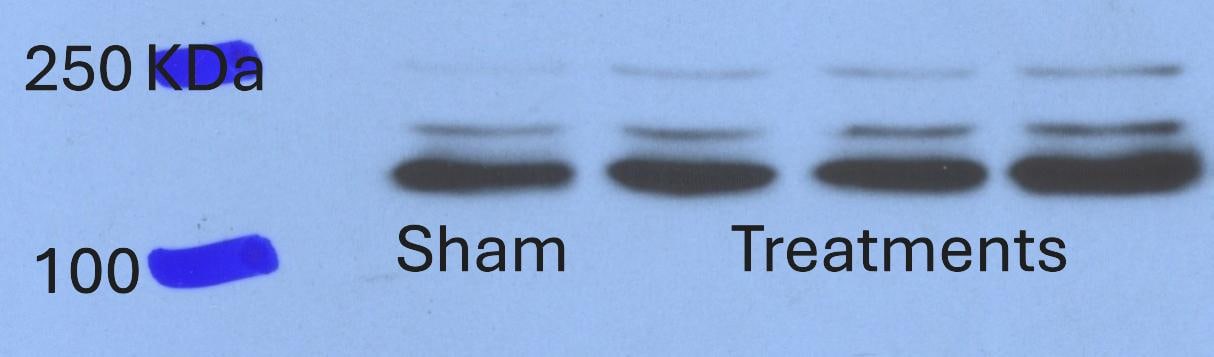
VEGFR3 (Flt-4), together with VEGFR1 (Flt-1) and VEGFR2 (KDR/Flk-1), belong to the class III subfamily of receptor tyrosine kinases (RTKs). All three receptors contain seven immunoglobulin-like repeats in their extracellular domains and kinase insert domains in their intracellular regions. The expression of these receptors is almost exclusively restricted to the endothelial cells. These receptors are likely to play essential roles in vasculogenesis and angiogenesis.

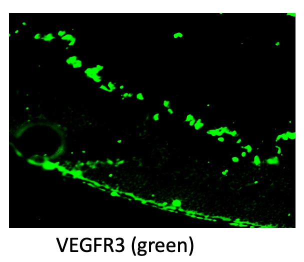
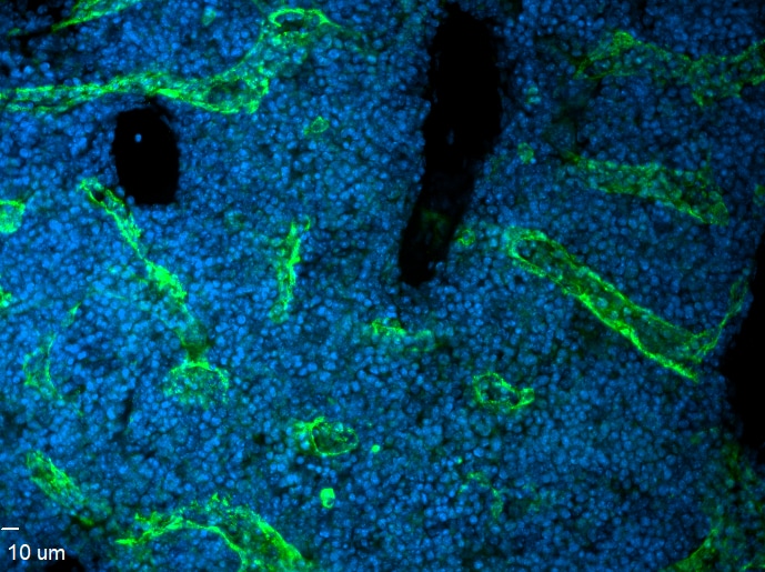
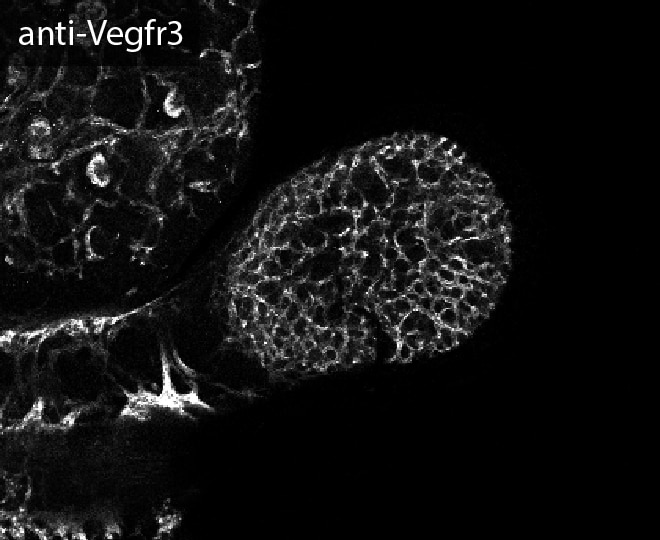
In adults, VEGFR3 expression is restricted to the endothelial cells of the lymphatic vessels. Mouse VEGFR3 cDNA encodes a 1363 amino acid (aa) residue precursor protein with a 24 aa residue signal peptide. Mature VEGFR3 has a 751 aa residue extracellular domain, a 22 aa residue hydrophobic transmembrane domain and a 565 aa residue cytoplasmic domain. The polypeptide sequences of murine Flt-4 is 88% identical to the human homologue. VEGFR3 has been reported to serve as the receptors for VEGF-C and VEGF-D.

References

  1. Finnerty, H. et al. (1993) Oncogene 8:2293.
  2. Joukov, V. et al. (1996) EMBO J. 15:290.
  3. Achen, M. et al. (1998) Proc. Natl. Acad. Sci. USA 95:548.

Long Name

Vascular Endothelial Growth Factor Receptor 3

Alternate Names

Flt-4, FLT4, VEGF R3

Entrez Gene IDs

2324 (Human); 14257 (Mouse)

Gene Symbol

FLT4

UniProt

Additional VEGFR3/Flt-4 Products

Product Documents for Mouse VEGFR3/Flt-4 Antibody

Certificate of Analysis

To download a Certificate of Analysis, please enter a lot or batch number in the search box below.

Note: Certificate of Analysis not available for kit components.

Product Specific Notices for Mouse VEGFR3/Flt-4 Antibody

For research use only

Citations for Mouse VEGFR3/Flt-4 Antibody

Customer Reviews for Mouse VEGFR3/Flt-4 Antibody (8)

4.4 out of 5
8 Customer Ratings
5 Stars
50%
4 Stars
38%
3 Stars
13%
2 Stars
0%
1 Stars
0%

Have you used Mouse VEGFR3/Flt-4 Antibody?

Submit a review and receive an Amazon gift card!

$25/€18/£15/$25CAN/¥2500 Yen for a review with an image

$10/€7/£6/$10CAN/¥1110 Yen for a review without an image

Submit a review
Amazon Gift Card

Customer Images


Showing  1 - 5 of 8 reviews Showing All
Filter By:
  • Mouse VEGFR3/Flt-4 Antibody
    Name: Anonymous
    Application: Western Blot
    Sample Tested: SVEC4-10 mouse vascular endothelial cell line
    Species: Mouse
    Verified Customer | Posted 11/13/2024
    Mouse VEGFR3/Flt-4 Antibody AF743
  • Mouse VEGFR3/Flt-4 Antibody
    Name: Anonymous
    Application: Immunocytochemistry/Immunofluorescence
    Sample Tested: Retina (outer nuclear layer)
    Species: Mouse
    Verified Customer | Posted 07/25/2023
    Mouse VEGFR3/Flt-4 Antibody AF743
  • Mouse VEGFR3/Flt-4 Antibody
    Name: Justin Vercellino
    Application: Immunocytochemistry/Immunofluorescence
    Sample Tested: bone marrow
    Species: Mouse
    Verified Customer | Posted 11/03/2020
    Whole-mounted mouse femur 5 ug of Ab used stained for 3 days at room temperature
    Mouse VEGFR3/Flt-4 Antibody AF743
  • Mouse VEGF R3/Flt-4 Antibody
    Name: Anonymous
    Application: Immunocytochemistry/Immunofluorescence
    Sample Tested: Adrenal gland tissue
    Species: Mouse
    Verified Customer | Posted 08/10/2017
    Mouse VEGFR3/Flt-4 Antibody AF743
  • Mouse VEGF R3/Flt-4 Antibody
    Name: Anonymous
    Application: Immunocytochemistry/Immunofluorescence
    Sample Tested: Mesentery whole mount
    Species: Mouse
    Verified Customer | Posted 10/04/2016
    Whole mount immunofluorescence of mouse mesenteric lymphatics. Mesentery was dissected from a P5 neonate, fixed in 2% PFA and stained using AF743 at a 1:200 dilution.
    Mouse VEGFR3/Flt-4 Antibody AF743
  • Name: Anonymous
    Application: Western Blot
    Sample Tested: See PMID 19901262
    Species: Mouse
    Verified Customer | Posted 01/09/2015
  • Name: Anonymous
    Application: Immunohistochemistry-Frozen
    Sample Tested: See PMID 23878309
    Species: Mouse
    Verified Customer | Posted 01/09/2015
  • Name: Anonymous
    Application: Immunofluorescence
    Sample Tested: See PMID 19901262
    Species: Mouse
    Verified Customer | Posted 01/09/2015

There are no reviews that match your criteria.

Showing  1 - 5 of 8 reviews Showing All

FAQs

No product specific FAQs exist for this product.

View all FAQs for Antibodies